6IK9

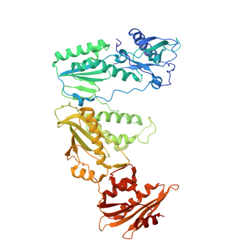
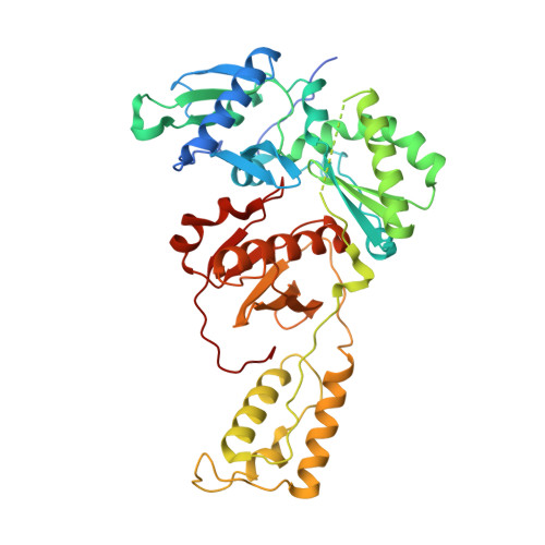
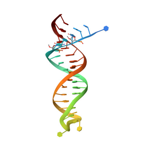
HIV-1 reverse transcriptase with Q151M/G112S/D113A/Y115F/F116Y/F160L/I159L:DNA:dGTP ternary complex


Experimental Data Snapshot

  • Method: X-RAY DIFFRACTION
  • Resolution: 2.44 Å
  • R-Value Free: 0.225 
  • R-Value Work: 0.186 
  • R-Value Observed: 0.188 

Starting Model: experimental
View more details

wwPDB Validation   3D Report Full Report


Ligand Structure Quality Assessment 


This is version 1.2 of the entry. See complete history


Literature

Active-site deformation in the structure of HIV-1 RT with HBV-associated septuple amino acid substitutions rationalizes the differential susceptibility of HIV-1 and HBV against 4'-modified nucleoside RT inhibitors.

Yasutake, Y.Hattori, S.I.Tamura, N.Matsuda, K.Kohgo, S.Maeda, K.Mitsuya, H.

(2019) Biochem Biophys Res Commun 509: 943-948

  • DOI: https://doi.org/10.1016/j.bbrc.2019.01.026
  • Primary Citation of Related Structures:  
    6IK9, 6IKA

  • PubMed Abstract: 

    Nucleoside analogue reverse transcriptase (RT) inhibitors (NRTIs) are major antiviral agents against hepatitis B virus (HBV) and human immunodeficiency virus type-1 (HIV-1). However, the notorious insoluble property of HBV RT has prevented atomic-resolution structural studies and rational anti-HBV drug design. Here, we created HIV-1 RT mutants containing HBV-mimicking sextuple or septuple amino acid substitutions at the nucleoside-binding site (N-site) and verified that these mutants retained the RT activity. The most active RT mutant, HIV-1 RT 7MC , carrying Q151M/G112S/D113A/Y115F/F116Y/F160L/I159L was successfully crystallized, and its three-dimensional structure was determined in complex with DNA:dGTP/entecavir-triphosphate (ETV-TP), a potent anti-HBV guanosine analogue RT inhibitor, at a resolution of 2.43 Å and 2.60 Å, respectively. The structures reveal significant positional rearrangements of the amino acid side-chains at the N-site, elucidating the mechanism underlying the differential susceptibility of HIV-1 and HBV against recently reported 4'-modified NRTIs.


  • Organizational Affiliation

    Bioproduction Research Institute, National Institute of Advanced Industrial Science and Technology (AIST), Sapporo, 062-8517, Japan; Computational Bio Big-Data Open Innovation Laboratory (CBBD-OIL), AIST, Sapporo, 062-8517, Japan. Electronic address: y-yasutake@aist.go.jp.


Macromolecules

Find similar proteins by:  (by identity cutoff)  |  3D Structure
Entity ID: 1
MoleculeChains Sequence LengthOrganismDetailsImage
HIV-1 reverse transcriptase p66 subunitA,
D [auth C]
557Human immunodeficiency virus 1Mutation(s): 9 
Gene Names: pol
UniProt
Find proteins for D3XFN7 (Human immunodeficiency virus type 1)
Explore D3XFN7 
Go to UniProtKB:  D3XFN7
Entity Groups  
Sequence Clusters30% Identity50% Identity70% Identity90% Identity95% Identity100% Identity
UniProt GroupD3XFN7
Sequence Annotations
Expand
  • Reference Sequence
Find similar proteins by:  (by identity cutoff)  |  3D Structure
Entity ID: 2
MoleculeChains Sequence LengthOrganismDetailsImage
HIV-1 reverse transcriptase p51 subunitB,
E [auth D]
444Human immunodeficiency virus 1Mutation(s): 2 
UniProt
Find proteins for P12497 (Human immunodeficiency virus type 1 group M subtype B (isolate NY5))
Explore P12497 
Go to UniProtKB:  P12497
Entity Groups  
Sequence Clusters30% Identity50% Identity70% Identity90% Identity95% Identity100% Identity
UniProt GroupP12497
Sequence Annotations
Expand
  • Reference Sequence
Find similar nucleic acids by:  (by identity cutoff)  |  3D Structure
Entity ID: 3
MoleculeChains LengthOrganismImage
DNA/RNA (38-MER)C [auth E],
F
38synthetic construct
Sequence Annotations
Expand
  • Reference Sequence
Small Molecules
Ligands 3 Unique
IDChains Name / Formula / InChI Key2D Diagram3D Interactions
DGT
Query on DGT

Download Ideal Coordinates CCD File 
G [auth A],
N [auth F]
2'-DEOXYGUANOSINE-5'-TRIPHOSPHATE
C10 H16 N5 O13 P3
HAAZLUGHYHWQIW-KVQBGUIXSA-N
GOL
Query on GOL

Download Ideal Coordinates CCD File 
I [auth B],
J [auth B],
L [auth D],
M [auth D]
GLYCEROL
C3 H8 O3
PEDCQBHIVMGVHV-UHFFFAOYSA-N
MG
Query on MG

Download Ideal Coordinates CCD File 
H [auth A],
K [auth C]
MAGNESIUM ION
Mg
JLVVSXFLKOJNIY-UHFFFAOYSA-N
Experimental Data & Validation

Experimental Data

  • Method: X-RAY DIFFRACTION
  • Resolution: 2.44 Å
  • R-Value Free: 0.225 
  • R-Value Work: 0.186 
  • R-Value Observed: 0.188 
  • Space Group: H 3
Unit Cell:
Length ( Å )Angle ( ˚ )
a = 284.869α = 90
b = 284.869β = 90
c = 95.892γ = 120
Software Package:
Software NamePurpose
PHENIXrefinement
XDSdata reduction
XDSdata scaling
PHENIXphasing

Structure Validation

View Full Validation Report


Ligand Structure Quality Assessment 


Entry History & Funding Information

Deposition Data


Funding OrganizationLocationGrant Number
Japan Agency for Medical Research and Development (AMED)JapanJP18fk0310113

Revision History  (Full details and data files)

  • Version 1.0: 2019-01-30
    Type: Initial release
  • Version 1.1: 2019-02-06
    Changes: Data collection, Database references
  • Version 1.2: 2023-11-22
    Changes: Data collection, Database references, Derived calculations, Refinement description