6IJM | pdb_00006ijm

Apo structure of the N6-methyl-AMP Deaminase from Arabidopsis thaliana


Experimental Data Snapshot

  • Method: X-RAY DIFFRACTION
  • Resolution: 2.02 Å
  • R-Value Free: 
    0.222 (Depositor), 0.228 (DCC) 
  • R-Value Work: 
    0.174 (Depositor), 0.181 (DCC) 
  • R-Value Observed: 
    0.177 (Depositor) 

Starting Model: experimental
View more details

wwPDB Validation 3D Report Full Report

Validation slider image for 6IJM

This is version 1.3 of the entry. See complete history

Literature

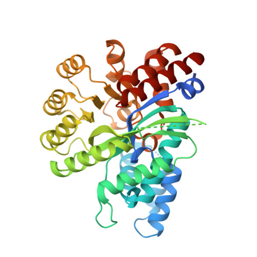
Alternative conformation induced by substrate binding for Arabidopsis thalianaN6-methyl-AMP deaminase.

Jia, Q.Xie, W.

(2019) Nucleic Acids Res 47: 3233-3243

  • DOI: https://doi.org/10.1093/nar/gkz070
  • Primary Citation Related Structures: 
    6IJM, 6IJN, 6IJP

  • PubMed Abstract: 

    Adenosine deaminase is involved in adenosine degradation and salvage pathway, and plays important physiological roles in purine metabolism. Recently, a novel type of adenosine deaminase-like protein has been identified, which displays deamination activity toward N6-methyl-adenosine monophosphate but not adenosine or AMP, and was consequently named N6-methyl-AMP deaminase (MAPDA). The underlying structural basis of MAPDA recognition and catalysis is poorly understood. Here, we present the crystal structures of MAPDA from Arabidopsis thaliana in the free and in the ligand-bound forms. The protein contains a conserved (β/α)8 Tim-barrel domain and a typical zinc-binding site, but it also exhibits idiosyncratic local differences for two flexible helices important for substrate binding. The extensive interactions between the N6-methyl-AMP substrate or the inosine monophosphate product and the enzyme were identified, and subsequently evaluated by the deamination activity assays. Importantly, each structure reported here represents a different stage of the catalytic pathway and their structural differences suggested that the enzyme can exist in two distinct conformational states. The open state switches to the closed one upon the binding of ligands, brought about by the two critical helices. Our structural studies provide the first look of this important metabolic enzyme and shed lights on its catalytic pathway, which holds promise for the structure-based drug design for MAPDA-related diseases.


  • Organizational Affiliation
    • MOE Key Laboratory of Gene Function and Regulation, State Key Laboratory for Biocontrol, School of Life Sciences, The Sun Yat-Sen University, Guangzhou, Guangdong, 510006, People's Republic of China.

Macromolecule Content 

  • Total Structure Weight: 42.56 kDa 
  • Atom Count: 2,807 
  • Modeled Residue Count: 337 
  • Deposited Residue Count: 376 
  • Unique protein chains: 1

Macromolecules

Find similar proteins by:|  3D Structure
Entity ID: 1
MoleculeChains  Sequence LengthOrganismDetailsImage
Adenosine/AMP deaminase family protein376Arabidopsis thalianaMutation(s): 0 
Gene Names: At4g04880T4B21.20T4B21_20
EC: 3.5.4
UniProt
Find proteins for Q8LPL7 (Arabidopsis thaliana)
Explore Q8LPL7 
Go to UniProtKB:  Q8LPL7
Entity Groups
Sequence Clusters30% Identity50% Identity70% Identity90% Identity95% Identity100% Identity
UniProt GroupQ8LPL7
Sequence Annotations
Expand
Reference Sequence

Small Molecules

Experimental Data & Validation

Experimental Data

  • Method: X-RAY DIFFRACTION
  • Resolution: 2.02 Å
  • R-Value Free:  0.222 (Depositor), 0.228 (DCC) 
  • R-Value Work:  0.174 (Depositor), 0.181 (DCC) 
  • R-Value Observed: 0.177 (Depositor) 
Space Group: P 21 21 21
Unit Cell:
Length ( Å )Angle ( ˚ )
a = 50.625α = 90
b = 79.862β = 90
c = 86.853γ = 90
Software Package:
Software NamePurpose
PHENIXrefinement
HKL-3000data reduction
HKL-3000data scaling
PHASERphasing

Structure Validation

View Full Validation Report


Entry History 

& Funding Information

Deposition Data


Funding OrganizationLocationGrant Number
National Natural Science Foundation of ChinaChina31870782
National Natural Science Foundation of ChinaChina31700657

Revision History  (Full details and data files)

  • Version 1.0: 2019-02-06
    Type: Initial release
  • Version 1.1: 2019-02-20
    Changes: Data collection, Database references
  • Version 1.2: 2019-04-17
    Changes: Data collection, Database references
  • Version 1.3: 2023-11-22
    Changes: Data collection, Database references, Refinement description