


Crystal structure of hypoxanthine phosphoribosyltransferase from Francisella tularensis
Pavithra, G.C., Ramagopal, U.A.To be published.
Experimental Data Snapshot
Starting Model: experimental
View more details
Entity ID: 1 | |||||
|---|---|---|---|---|---|
| Molecule | Chains | Sequence Length | Organism | Details | Image |
| Hypoxanthine phosphoribosyltransferase | 177 | Francisella tularensis | Mutation(s): 0 Gene Names: hpt EC: 2.4.2.8 | 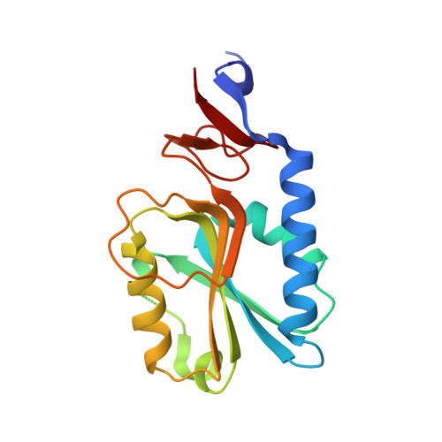 | |
UniProt | |||||
Entity Groups | |||||
| Sequence Clusters | 30% Identity50% Identity70% Identity90% Identity95% Identity100% Identity | ||||
| UniProt Group | Q5NI77 | ||||
Sequence AnnotationsExpand | |||||
Reference Sequence | |||||
| Ligands 2 Unique | |||||
|---|---|---|---|---|---|
| ID | Chains | Name / Formula / InChI Key | 2D Diagram | 3D Interactions | |
| SO4 (Subject of Investigation/LOI) Download:Ideal Coordinates CCD File | E [auth A] F [auth A] I [auth B] L [auth C] M [auth C] | SULFATE ION O4 S QAOWNCQODCNURD-UHFFFAOYSA-L |  | ||
| ZN (Subject of Investigation/LOI) Download:Ideal Coordinates CCD File | G [auth A] H [auth A] J [auth B] K [auth B] N [auth C] | ZINC ION Zn PTFCDOFLOPIGGS-UHFFFAOYSA-N |  | ||
| Length ( Å ) | Angle ( ˚ ) |
|---|---|
| a = 78.764 | α = 90 |
| b = 90.193 | β = 90 |
| c = 113.707 | γ = 90 |
| Software Name | Purpose |
|---|---|
| SCALEPACK | data scaling |
| REFMAC | refinement |
| PDB_EXTRACT | data extraction |
| DENZO | data reduction |
| MOLREP | phasing |


