6IDG | pdb_00006idg

antibody 64M-5 Fab in complex with dT(6-4)T


Experimental Data Snapshot

  • Method: X-RAY DIFFRACTION
  • Resolution: 2.00 Å
  • R-Value Free: 
    0.240 (Depositor), 0.239 (DCC) 
  • R-Value Work: 
    0.192 (Depositor), 0.192 (DCC) 
  • R-Value Observed: 
    0.192 (Depositor) 

Starting Model: experimental
View more details

wwPDB Validation 3D Report Full Report

Validation slider image for 6IDG

This is version 1.3 of the entry. See complete history

Literature

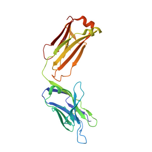
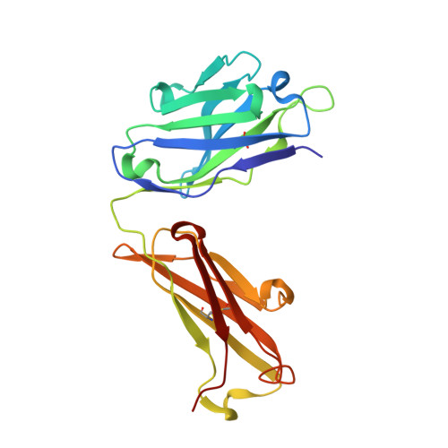
Structures of the antibody 64M-5 Fab and its complex with dT(6-4)T indicate induced-fit and high-affinity mechanisms.

Yokoyama, H.Mizutani, R.Noguchi, S.Hayashida, N.

(2019) Acta Crystallogr F Struct Biol Commun 75: 80-88

  • DOI: https://doi.org/10.1107/S2053230X18017661
  • Primary Citation Related Structures: 
    6IDG, 6IDH

  • PubMed Abstract: 

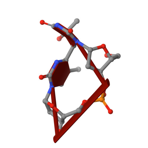
    DNA photoproducts with (6-4) pyrimidine-pyrimidone adducts produced by ultraviolet light are mutagenic and carcinogenic. The crystal structures of the anti-(6-4) photoproduct antibody 64M-5 Fab and of its complex with dT(6-4)T were determined at 2.5 and 2.0 Å resolution, respectively. A comparison between the dT(6-4)T-liganded and unliganded structures indicates that the side chain of His93L is greatly rotated and shifted on binding to dT(6-4)T, leading to the formation of an electrostatic interaction with the phosphate moiety of dT(6-4)T, which shows a remarkable induced fit. Based on a comparison of the dT(6-4)T-liganded structures of the 64M-5 and 64M-2 Fabs, the electrostatic interaction between the side chain of His93L in 64M-5 and the phosphate moiety of dT(6-4)T is lost for Leu93L in 64M-2, while Arg90L in 64M-5 instead of Gln90L in 64M-2 stabilizes the conformation of complementarity-determining region (CDR) L3. These differences contribute to the higher affinity of 64M-5 for dT(6-4)T compared with that of 64M-2.


  • Organizational Affiliation
    • Faculty of Pharmaceutical Sciences, Tokyo University of Science, 2641 Yamazaki, Noda, Chiba 278-8510, Japan.

Macromolecule Content 

  • Total Structure Weight: 48.39 kDa 
  • Atom Count: 3,780 
  • Modeled Residue Count: 440 
  • Deposited Residue Count: 440 
  • Unique protein chains: 2
  • Unique nucleic acid chains: 1

Macromolecules


Find similar proteins by:|  3D Structure
Entity ID: 1
MoleculeChains  Sequence LengthOrganismDetailsImage
Anti-(6-4) photoproduct antibody 64M-5 Fab (light chain)A [auth L]217Mus musculusMutation(s): 0 
Find similar proteins by:|  3D Structure
Entity ID: 2
MoleculeChains  Sequence LengthOrganismDetailsImage
Anti-(6-4) photoproduct antibody 64M-5 Fab (heavy chain)B [auth H]221Mus musculusMutation(s): 0 
Find similar nucleic acids by:  Sequence
Entity ID: 3
MoleculeChains LengthOrganismImage
DNA (5'-D(*(64T)P*(5PY))-3')C [auth A]2synthetic construct
Sequence Annotations
Expand
Reference Sequence

Experimental Data & Validation

Experimental Data

  • Method: X-RAY DIFFRACTION
  • Resolution: 2.00 Å
  • R-Value Free:  0.240 (Depositor), 0.239 (DCC) 
  • R-Value Work:  0.192 (Depositor), 0.192 (DCC) 
  • R-Value Observed: 0.192 (Depositor) 
Space Group: P 21 21 21
Unit Cell:
Length ( Å )Angle ( ˚ )
a = 84.62α = 90
b = 100.865β = 90
c = 51.666γ = 90
Software Package:
Software NamePurpose
CNSrefinement
DENZOdata reduction
SCALEPACKdata scaling
X-PLORphasing

Structure Validation

View Full Validation Report


Entry History 

& Funding Information

Deposition Data


Funding OrganizationLocationGrant Number
Japan Society for the Promotion of ScienceJapan17K07316

Revision History  (Full details and data files)

  • Version 1.0: 2019-02-13
    Type: Initial release
  • Version 1.1: 2019-11-20
    Changes: Database references
  • Version 1.2: 2023-11-22
    Changes: Data collection, Database references, Refinement description
  • Version 1.3: 2024-10-09
    Changes: Structure summary