6IAT

Icosahedrally averaged capsid of bacteriophage P68


Experimental Data Snapshot

  • Method: ELECTRON MICROSCOPY
  • Resolution: 3.30 Å
  • Aggregation State: PARTICLE 
  • Reconstruction Method: SINGLE PARTICLE 

wwPDB Validation   3D Report Full Report


This is version 1.2 of the entry. See complete history


Literature

Structure and genome ejection mechanism ofStaphylococcus aureusphage P68.

Hrebik, D.Stverakova, D.Skubnik, K.Fuzik, T.Pantucek, R.Plevka, P.

(2019) Sci Adv 5: eaaw7414-eaaw7414

  • DOI: https://doi.org/10.1126/sciadv.aaw7414
  • Primary Citation of Related Structures:  
    6IAB, 6IAC, 6IAT, 6IAW, 6IB1, 6Q3G

  • PubMed Abstract: 

    Phages infecting Staphylococcus aureus can be used as therapeutics against antibiotic-resistant bacterial infections. However, there is limited information about the mechanism of genome delivery of phages that infect Gram-positive bacteria. Here, we present the structures of native S. aureus phage P68, genome ejection intermediate, and empty particle. The P68 head contains 72 subunits of inner core protein, 15 of which bind to and alter the structure of adjacent major capsid proteins and thus specify attachment sites for head fibers. Unlike in the previously studied phages, the head fibers of P68 enable its virion to position itself at the cell surface for genome delivery. The unique interaction of one end of P68 DNA with one of the 12 portal protein subunits is disrupted before the genome ejection. The inner core proteins are released together with the DNA and enable the translocation of phage genome across the bacterial membrane into the cytoplasm.


  • Organizational Affiliation

    Central European Institute of Technology, Masaryk University, Kamenice 5, 625 00 Brno, Czech Republic.


Macromolecules
Find similar proteins by:  (by identity cutoff)  |  3D Structure
Entity ID: 1
MoleculeChains Sequence LengthOrganismDetailsImage
Major head proteinA [auth C],
B [auth A],
C [auth B],
D
408Staphylococcus phage P68Mutation(s): 0 
UniProt
Find proteins for Q859I3 (Staphylococcus phage P68)
Explore Q859I3 
Go to UniProtKB:  Q859I3
Entity Groups  
Sequence Clusters30% Identity50% Identity70% Identity90% Identity95% Identity100% Identity
UniProt GroupQ859I3
Sequence Annotations
Expand
  • Reference Sequence
Find similar proteins by:  (by identity cutoff)  |  3D Structure
Entity ID: 2
MoleculeChains Sequence LengthOrganismDetailsImage
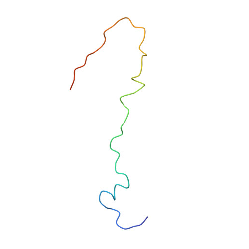
Arstotzka protein
E, F, G, H
60Staphylococcus phage P68Mutation(s): 0 
UniProt
Find proteins for Q859I2 (Staphylococcus phage P68)
Explore Q859I2 
Go to UniProtKB:  Q859I2
Entity Groups  
Sequence Clusters30% Identity50% Identity70% Identity90% Identity95% Identity100% Identity
UniProt GroupQ859I2
Sequence Annotations
Expand
  • Reference Sequence
Experimental Data & Validation

Experimental Data

  • Method: ELECTRON MICROSCOPY
  • Resolution: 3.30 Å
  • Aggregation State: PARTICLE 
  • Reconstruction Method: SINGLE PARTICLE 
EM Software:
TaskSoftware PackageVersion
MODEL REFINEMENTPHENIXdev:3042
RECONSTRUCTIONRELION2.1

Structure Validation

View Full Validation Report


Entry History & Funding Information

Deposition Data


Funding OrganizationLocationGrant Number
Czech Science FoundationCzech Republic15-21631Y
Czech Science FoundationCzech Republic18-17810S
European Molecular Biology OrganizationCzech Republic3041
Czech Republic16-29916A

Revision History  (Full details and data files)

  • Version 1.0: 2019-11-06
    Type: Initial release
  • Version 1.1: 2019-11-13
    Changes: Data collection, Other
  • Version 1.2: 2024-05-15
    Changes: Data collection, Database references, Derived calculations