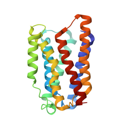
6I6B

Crystal structure of the KDEL receptor in the peptide free state.


Experimental Data Snapshot

  • Method: X-RAY DIFFRACTION
  • Resolution: 2.59 Å
  • R-Value Free: 0.271 
  • R-Value Work: 0.242 
  • R-Value Observed: 0.244 

Starting Model: other
View more details

wwPDB Validation   3D Report Full Report


Ligand Structure Quality Assessment 


This is version 1.3 of the entry. See complete history


Literature

Structural basis for pH-dependent retrieval of ER proteins from the Golgi by the KDEL receptor.

Brauer, P.Parker, J.L.Gerondopoulos, A.Zimmermann, I.Seeger, M.A.Barr, F.A.Newstead, S.

(2019) Science 363: 1103-1107

  • DOI: https://doi.org/10.1126/science.aaw2859
  • Primary Citation of Related Structures:  
    6I6B, 6I6H, 6I6J

  • PubMed Abstract: 

    Selective export and retrieval of proteins between the endoplasmic reticulum (ER) and Golgi apparatus is indispensable for eukaryotic cell function. An essential step in the retrieval of ER luminal proteins from the Golgi is the pH-dependent recognition of a carboxyl-terminal Lys-Asp-Glu-Leu (KDEL) signal by the KDEL receptor. Here, we present crystal structures of the chicken KDEL receptor in the apo ER state, KDEL-bound Golgi state, and in complex with an antagonistic synthetic nanobody (sybody). These structures show a transporter-like architecture that undergoes conformational changes upon KDEL binding and reveal a pH-dependent interaction network crucial for recognition of the carboxyl terminus of the KDEL signal. Complementary in vitro binding and in vivo cell localization data explain how these features create a pH-dependent retrieval system in the secretory pathway.


  • Organizational Affiliation

    Department of Biochemistry, University of Oxford, South Parks Road, Oxford OX1 3QU, UK.


Macromolecules
Find similar proteins by:  (by identity cutoff)  |  3D Structure
Entity ID: 1
MoleculeChains Sequence LengthOrganismDetailsImage
ER lumen protein-retaining receptor 2203Gallus gallusMutation(s): 0 
Gene Names: KDELR2RCJMB04_8l23
Membrane Entity: Yes 
UniProt
Find proteins for Q5ZKX9 (Gallus gallus)
Explore Q5ZKX9 
Go to UniProtKB:  Q5ZKX9
Entity Groups  
Sequence Clusters30% Identity50% Identity70% Identity90% Identity95% Identity100% Identity
UniProt GroupQ5ZKX9
Sequence Annotations
Expand
  • Reference Sequence
Experimental Data & Validation

Experimental Data

  • Method: X-RAY DIFFRACTION
  • Resolution: 2.59 Å
  • R-Value Free: 0.271 
  • R-Value Work: 0.242 
  • R-Value Observed: 0.244 
  • Space Group: C 2 2 21
Unit Cell:
Length ( Å )Angle ( ˚ )
a = 47.899α = 90
b = 103.75β = 90
c = 101.588γ = 90
Software Package:
Software NamePurpose
PHENIXrefinement
xia2data scaling
PDB_EXTRACTdata extraction
DIALSdata reduction
PHASERphasing

Structure Validation

View Full Validation Report


Ligand Structure Quality Assessment 


Entry History & Funding Information

Deposition Data


Funding OrganizationLocationGrant Number
Wellcome TrustUnited Kingdom109133/Z/15/A

Revision History  (Full details and data files)

  • Version 1.0: 2019-02-27
    Type: Initial release
  • Version 1.1: 2019-03-06
    Changes: Data collection, Database references
  • Version 1.2: 2019-03-20
    Changes: Data collection, Database references
  • Version 1.3: 2024-05-01
    Changes: Data collection, Database references, Refinement description