6I5M | pdb_00006i5m

Gamma subunit of the translation initiation factor 2 from Sulfolobus solfataricus in complex with GDP and formate ion


Experimental Data Snapshot

  • Method: X-RAY DIFFRACTION
  • Resolution: 2.40 Å
  • R-Value Free: 
    0.224 (Depositor), 0.221 (DCC) 
  • R-Value Work: 
    0.173 (Depositor), 0.174 (DCC) 
  • R-Value Observed: 
    0.175 (Depositor) 

Starting Model: experimental
View more details

wwPDB Validation 3D Report Full Report

Validation slider image for 6I5M

Ligand Structure Quality Assessment 


This is version 1.3 of the entry. See complete history

Literature

The third structural switch in the archaeal translation initiation factor 2 (aIF2) molecule and its possible role in the initiation of GTP hydrolysis and the removal of aIF2 from the ribosome.

Nikonov, O.Kravchenko, O.Nevskaya, N.Stolboushkina, E.Garber, M.Nikonov, S.

(2019) Acta Crystallogr D Struct Biol 75: 392-399

  • DOI: https://doi.org/10.1107/S2059798319002304
  • Primary Citation Related Structures: 
    6H6K, 6I5M

  • PubMed Abstract: 

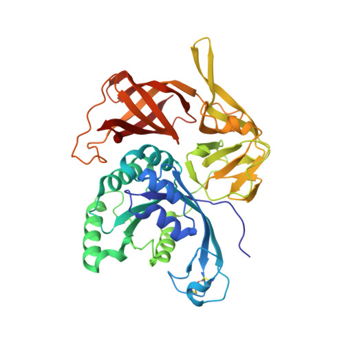
    The structure of the γ subunit of archaeal translation initiation factor 2 (aIF2) from Sulfolobus solfataricus (SsoIF2γ) was determined in complex with GDPCP (a GTP analog). Crystals were obtained in the absence of magnesium ions in the crystallization solution. They belonged to space group P1, with five molecules in the unit cell. Four of these molecules are related in pairs by a common noncrystallographic twofold symmetry axis, while the fifth has no symmetry equivalent. Analysis of the structure and its comparison with other known aIF2 γ-subunit structures in the GTP-bound state show that (i) the magnesium ion is necessary for the formation and the maintenance of the active form of SsoIF2γ and (ii) in addition to the two previously known structural switches 1 and 2, eukaryotic translation initiation factor 2 (eIF2) and aIF2 molecules have another flexible region (switch 3), the function of which may consist of initiation of the hydrolysis of GTP and the removal of e/aIF2 from the ribosome after codon-anticodon recognition.


  • Organizational Affiliation
    • Institute of Protein Research, Russian Academy of Sciences, 142290 Pushchino, Russian Federation.

Macromolecule Content 

  • Total Structure Weight: 47.15 kDa 
  • Atom Count: 3,744 
  • Modeled Residue Count: 414 
  • Deposited Residue Count: 415 
  • Unique protein chains: 1

Macromolecules

Find similar proteins by:|  3D Structure
Entity ID: 1
MoleculeChains  Sequence LengthOrganismDetailsImage
Translation initiation factor 2 subunit gamma415Saccharolobus solfataricusMutation(s): 0 
Gene Names: eif2gSSO0412
EC: 3.6.5.3
UniProt
Find proteins for Q980A5 (Saccharolobus solfataricus (strain ATCC 35092 / DSM 1617 / JCM 11322 / P2))
Explore Q980A5 
Go to UniProtKB:  Q980A5
Entity Groups
Sequence Clusters30% Identity50% Identity70% Identity90% Identity95% Identity100% Identity
UniProt GroupQ980A5
Sequence Annotations
Expand
Reference Sequence

Small Molecules

Ligands 4 Unique
IDChains Name / Formula / InChI Key2D Diagram3D Interactions
GDP

Query on GDP


Download:Ideal Coordinates CCD File
B [auth A]GUANOSINE-5'-DIPHOSPHATE
C10 H15 N5 O11 P2
QGWNDRXFNXRZMB-UUOKFMHZSA-N
FMT

Query on FMT


Download:Ideal Coordinates CCD File
C [auth A]
D [auth A]
E [auth A]
F [auth A]
G [auth A]
C [auth A],
D [auth A],
E [auth A],
F [auth A],
G [auth A],
H [auth A],
I [auth A],
J [auth A],
K [auth A],
L [auth A],
M [auth A],
N [auth A],
O [auth A],
P [auth A],
Q [auth A],
R [auth A],
S [auth A]
FORMIC ACID
C H2 O2
BDAGIHXWWSANSR-UHFFFAOYSA-N
MG

Query on MG


Download:Ideal Coordinates CCD File
T [auth A],
U [auth A]
MAGNESIUM ION
Mg
JLVVSXFLKOJNIY-UHFFFAOYSA-N
NA

Query on NA


Download:Ideal Coordinates CCD File
V [auth A]SODIUM ION
Na
FKNQFGJONOIPTF-UHFFFAOYSA-N

Experimental Data & Validation

Experimental Data

  • Method: X-RAY DIFFRACTION
  • Resolution: 2.40 Å
  • R-Value Free:  0.224 (Depositor), 0.221 (DCC) 
  • R-Value Work:  0.173 (Depositor), 0.174 (DCC) 
  • R-Value Observed: 0.175 (Depositor) 
Space Group: H 3 2
Unit Cell:
Length ( Å )Angle ( ˚ )
a = 142.143α = 90
b = 142.143β = 90
c = 218.587γ = 120
Software Package:
Software NamePurpose
PHENIXrefinement
PROTEUM PLUSdata reduction
PROTEUM PLUSdata scaling
PHASERphasing

Structure Validation

View Full Validation Report


Ligand Structure Quality Assessment 


Entry History 

Deposition Data

Revision History  (Full details and data files)

  • Version 1.0: 2019-01-02
    Type: Initial release
  • Version 1.1: 2019-04-24
    Changes: Data collection, Database references
  • Version 1.2: 2024-01-24
    Changes: Data collection, Database references, Derived calculations, Refinement description
  • Version 1.3: 2024-10-16
    Changes: Structure summary