6I4B | pdb_00006i4b

Plasmodium falciparum dihydroorotate dehydrogenase (DHODH) co-crystallized with 3-Hydroxy-1-methyl-5-((3-(trifluoromethyl)phenoxy)methyl)-1H-pyrazole-4-carboxylic acid


Experimental Data Snapshot

  • Method: X-RAY DIFFRACTION
  • Resolution: 1.98 Å
  • R-Value Free: 
    0.226 (Depositor), 0.227 (DCC) 
  • R-Value Work: 
    0.192 (Depositor), 0.194 (DCC) 
  • R-Value Observed: 
    0.194 (Depositor) 

wwPDB Validation 3D Report Full Report

Validation slider image for 6I4B

Ligand Structure Quality Assessment 


This is version 1.2 of the entry. See complete history

Literature

Hydroxyazole scaffold-based Plasmodium falciparum dihydroorotate dehydrogenase inhibitors: Synthesis, biological evaluation and X-ray structural studies.

Pippione, A.C.Sainas, S.Goyal, P.Fritzson, I.Cassiano, G.C.Giraudo, A.Giorgis, M.Tavella, T.A.Bagnati, R.Rolando, B.Caing-Carlsson, R.Costa, F.T.M.Andrade, C.H.Al-Karadaghi, S.Boschi, D.Friemann, R.Lolli, M.L.

(2018) Eur J Med Chem 163: 266-280

  • DOI: https://doi.org/10.1016/j.ejmech.2018.11.044
  • Primary Citation Related Structures: 
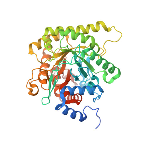
    6I4B, 6I55

  • PubMed Abstract: 

    Plasmodium falciparum dihydroorotate dehydrogenase (PfDHODH) has been clinically validated as a target for antimalarial drug discovery, as a triazolopyrimidine class inhibitor (DSM265) is currently undergoing clinical development. Here, we have identified new hydroxyazole scaffold-based PfDHODH inhibitors belonging to two different chemical series. The first series was designed by a scaffold hopping strategy that exploits the use of hydroxylated azoles. Within this series, the hydroxythiadiazole 3 was identified as the best selective PfDHODH inhibitor (IC 50 12.0 μM). The second series was designed by modulating four different positions of the hydroxypyrazole scaffold. In particular, hydroxypyrazoles 7e and 7f were shown to be active in the low μM range (IC 50 2.8 and 5.3 μM, respectively). All three compounds, 3, 7e and 7f showed clear selectivity over human DHODH (IC 50  > 200 μM), low cytotoxicity, and retained micromolar activity in P. falciparum-infected erythrocytes. The crystallographic structures of PfDHODH in complex with compounds 3 and 7e proved their binding mode, supplying essential data for future optimization of these scaffolds.


  • Organizational Affiliation
    • Department of Science and Drug Technology, University of Turin, via Pietro Giuria 9, 10125, Turin, Italy.

Macromolecule Content 

  • Total Structure Weight: 93.56 kDa 
  • Atom Count: 6,422 
  • Modeled Residue Count: 755 
  • Deposited Residue Count: 810 
  • Unique protein chains: 1

Macromolecules

Find similar proteins by:|  3D Structure
Entity ID: 1
MoleculeChains  Sequence LengthOrganismDetailsImage
Dihydroorotate dehydrogenase (quinone), mitochondrial
A, B
405Plasmodium falciparumMutation(s): 0 
Gene Names: PFF0160c
EC: 1.3.5.2
Membrane Entity: Yes 
UniProt
Find proteins for Q08210 (Plasmodium falciparum (isolate 3D7))
Explore Q08210 
Go to UniProtKB:  Q08210
Entity Groups
Sequence Clusters30% Identity50% Identity70% Identity90% Identity95% Identity100% Identity
UniProt GroupQ08210
Sequence Annotations
Expand
Reference Sequence

Small Molecules

Ligands 3 Unique
IDChains Name / Formula / InChI Key2D Diagram3D Interactions
FMN

Query on FMN


Download:Ideal Coordinates CCD File
C [auth A],
F [auth B]
FLAVIN MONONUCLEOTIDE
C17 H21 N4 O9 P
FVTCRASFADXXNN-SCRDCRAPSA-N
E2N
(Subject of Investigation/LOI)

Query on E2N


Download:Ideal Coordinates CCD File
E [auth A],
H [auth B]
1-methyl-3-oxidanyl-5-[[3-(trifluoromethyl)phenoxy]methyl]pyrazole-4-carboxylic acid
C13 H11 F3 N2 O4
ZOSMQYUVHWSYCZ-UHFFFAOYSA-N
ORO

Query on ORO


Download:Ideal Coordinates CCD File
D [auth A],
G [auth B]
OROTIC ACID
C5 H4 N2 O4
PXQPEWDEAKTCGB-UHFFFAOYSA-N
Binding Affinity Annotations 
IDSourceBinding Affinity
E2N BindingDB:  6I4B IC50: 2800 (nM) from 1 assay(s)

Experimental Data & Validation

Experimental Data

  • Method: X-RAY DIFFRACTION
  • Resolution: 1.98 Å
  • R-Value Free:  0.226 (Depositor), 0.227 (DCC) 
  • R-Value Work:  0.192 (Depositor), 0.194 (DCC) 
  • R-Value Observed: 0.194 (Depositor) 
Space Group: P 1 21 1
Unit Cell:
Length ( Å )Angle ( ˚ )
a = 50.607α = 90
b = 158.121β = 105.67
c = 61.753γ = 90
Software Package:
Software NamePurpose
XDSdata reduction
SCALAdata scaling
PHENIXrefinement
PDB_EXTRACTdata extraction
PHASERphasing

Structure Validation

View Full Validation Report


Ligand Structure Quality Assessment 


Entry History 

Deposition Data

Revision History  (Full details and data files)

  • Version 1.0: 2018-12-19
    Type: Initial release
  • Version 1.1: 2024-05-15
    Changes: Data collection, Database references
  • Version 1.2: 2025-12-24
    Changes: Database references, Source and taxonomy, Structure summary