6I1K | pdb_00006i1k

Crystal structure of catalytically inactive FnCas12a in complex with a crRNA guide and a dsDNA target


Experimental Data Snapshot

  • Method: X-RAY DIFFRACTION
  • Resolution: 2.65 Å
  • R-Value Free: 
    0.264 (Depositor), 0.267 (DCC) 
  • R-Value Work: 
    0.248 (Depositor), 0.250 (DCC) 
  • R-Value Observed: 
    0.249 (Depositor) 

Starting Model: experimental
View more details

wwPDB Validation 3D Report Full Report

Validation slider image for 6I1K

This is version 1.3 of the entry. See complete history

Literature

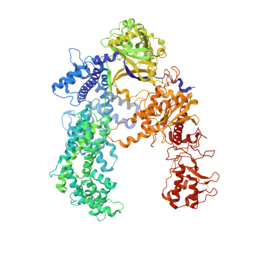
Mechanistic Insights into the cis- and trans-Acting DNase Activities of Cas12a.

Swarts, D.C.Jinek, M.

(2019) Mol Cell 73: 589-600.e4

  • DOI: https://doi.org/10.1016/j.molcel.2018.11.021
  • Primary Citation Related Structures: 
    6I1K, 6I1L

  • PubMed Abstract: 

    CRISPR-Cas12a (Cpf1) is an RNA-guided DNA-cutting nuclease that has been repurposed for genome editing. Upon target DNA binding, Cas12a cleaves both the target DNA in cis and non-target single-stranded DNAs (ssDNAs) in trans. To elucidate the molecular basis for both DNase cleavage modes, we performed structural and biochemical studies on Francisella novicida Cas12a. We show that guide RNA-target strand DNA hybridization conformationally activates Cas12a, triggering its trans-acting, non-specific, single-stranded DNase activity. In turn, cis cleavage of double-stranded DNA targets is a result of protospacer adjacent motif (PAM)-dependent DNA duplex unwinding, electrostatic stabilization of the displaced non-target DNA strand, and ordered sequential cleavage of the non-target and target DNA strands. Cas12a releases the PAM-distal DNA cleavage product and remains bound to the PAM-proximal DNA cleavage product in a catalytically competent, trans-active state. Together, these results provide a revised model for the molecular mechanisms of both the cis- and the trans-acting DNase activities of Cas12a enzymes, enabling their further exploitation as genome editing tools.


  • Organizational Affiliation
    • Department of Biochemistry, University of Zurich, 8057 Zurich, Switzerland.

Macromolecule Content 

  • Total Structure Weight: 189.46 kDa 
  • Atom Count: 12,982 
  • Modeled Residue Count: 1,393 
  • Deposited Residue Count: 1,418 
  • Unique protein chains: 1
  • Unique nucleic acid chains: 3

Macromolecules


Find similar proteins by:|  3D Structure
Entity ID: 1
MoleculeChains  Sequence LengthOrganismDetailsImage
CRISPR-associated endonuclease Cas12a1,302Francisella tularensis subsp. novicida U112Mutation(s): 0 
Gene Names: cas12acpf1FTN_1397
EC: 3.1.21.1 (PDB Primary Data), 3.1.27.2 (PDB Primary Data), 4.6.1.22 (UniProt)
UniProt
Find proteins for A0Q7Q2 (Francisella tularensis subsp. novicida (strain ATCC 15482 / CCUG 33449 / U112))
Explore A0Q7Q2 
Go to UniProtKB:  A0Q7Q2
Entity Groups
Sequence Clusters30% Identity50% Identity70% Identity90% Identity95% Identity100% Identity
UniProt GroupA0Q7Q2
Sequence Annotations
Expand
Reference Sequence
Find similar nucleic acids by:  (by identity cutoff) 
Entity ID: 2
MoleculeChains LengthOrganismImage
crRNA (40-MER)40synthetic construct
Sequence Annotations
Expand
Reference Sequence
Find similar nucleic acids by:  (by identity cutoff) 
Entity ID: 3
MoleculeChains LengthOrganismImage
DNA target strand (38-MER)38synthetic construct
Sequence Annotations
Expand
Reference Sequence
Find similar nucleic acids by:  (by identity cutoff) 
Entity ID: 4
MoleculeChains LengthOrganismImage
DNA non-target strand (33-MER)38synthetic construct
Sequence Annotations
Expand
Reference Sequence

Small Molecules

Ligands 2 Unique
IDChains Name / Formula / InChI Key2D Diagram3D Interactions
EDO

Query on EDO


Download:Ideal Coordinates CCD File
AA [auth C]
BA [auth C]
CA [auth C]
N [auth A]
O [auth A]
AA [auth C],
BA [auth C],
CA [auth C],
N [auth A],
O [auth A],
P [auth A],
Q [auth A],
R [auth A],
U [auth B],
V [auth B],
W [auth B],
X [auth B],
Y [auth B],
Z [auth B]
1,2-ETHANEDIOL
C2 H6 O2
LYCAIKOWRPUZTN-UHFFFAOYSA-N
MG

Query on MG


Download:Ideal Coordinates CCD File
E [auth A]
F [auth A]
G [auth A]
H [auth A]
I [auth A]
E [auth A],
F [auth A],
G [auth A],
H [auth A],
I [auth A],
J [auth A],
K [auth A],
L [auth A],
M [auth A],
S [auth B],
T [auth B]
MAGNESIUM ION
Mg
JLVVSXFLKOJNIY-UHFFFAOYSA-N

Experimental Data & Validation

Experimental Data

  • Method: X-RAY DIFFRACTION
  • Resolution: 2.65 Å
  • R-Value Free:  0.264 (Depositor), 0.267 (DCC) 
  • R-Value Work:  0.248 (Depositor), 0.250 (DCC) 
  • R-Value Observed: 0.249 (Depositor) 
Space Group: P 1 21 1
Unit Cell:
Length ( Å )Angle ( ˚ )
a = 82.172α = 90
b = 141.681β = 97.43
c = 89.478γ = 90
Software Package:
Software NamePurpose
PHENIXrefinement
XDSdata reduction
PHASERphasing
Cootmodel building
XSCALEdata scaling

Structure Validation

View Full Validation Report


Entry History 

& Funding Information

Deposition Data


Funding OrganizationLocationGrant Number
European Molecular Biology OrganizationGermanyALTF 179-2015
European Molecular Biology OrganizationGermanyaALTF 509-2017
Swiss National Science FoundationSwitzerlandSNSF 31003A_149393

Revision History  (Full details and data files)

  • Version 1.0: 2019-01-23
    Type: Initial release
  • Version 1.1: 2019-02-20
    Changes: Data collection, Database references
  • Version 1.2: 2024-01-24
    Changes: Data collection, Database references, Derived calculations, Refinement description
  • Version 1.3: 2024-10-09
    Changes: Structure summary