6I0R | pdb_00006i0r

Structure of quinolinate synthase in complex with 5-mercaptopyridine-2,3-dicarboxylic acid


Experimental Data Snapshot

  • Method: X-RAY DIFFRACTION
  • Resolution: 2.10 Å
  • R-Value Free: 
    0.239 (Depositor), 0.249 (DCC) 
  • R-Value Work: 
    0.198 (Depositor), 0.205 (DCC) 
  • R-Value Observed: 
    0.200 (Depositor) 

Starting Model: experimental
View more details

wwPDB Validation 3D Report Full Report

Validation slider image for 6I0R

Ligand Structure Quality Assessment 


This is version 1.2 of the entry. See complete history

Literature

Design of specific inhibitors of quinolinate synthase based on [4Fe-4S] cluster coordination.

Saez Cabodevilla, J.Volbeda, A.Hamelin, O.Latour, J.M.Gigarel, O.Clemancey, M.Darnault, C.Reichmann, D.Amara, P.Fontecilla-Camps, J.C.Ollagnier de Choudens, S.

(2019) Chem Commun (Camb) 55: 3725-3728

  • DOI: https://doi.org/10.1039/c8cc09023h
  • Primary Citation Related Structures: 
    6I0K, 6I0P, 6I0R

  • PubMed Abstract: 

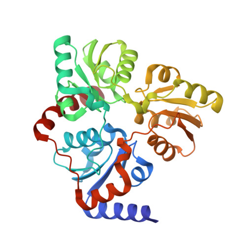
    Quinolinate synthase (NadA) is a [4Fe-4S] cluster-containing enzyme involved in the formation of quinolinic acid, the precursor of the essential NAD coenzyme. Here, we report the synthesis and activity of derivatives of the first inhibitor of NadA. Using multidisciplinary approaches we have investigated their action mechanism and discovered additional specific inhibitors of this enzyme.


  • Organizational Affiliation
    • Univ. Grenoble Alpes, CEA, CNRS, BIG-LCBM, UMR5249, 38000, Grenoble, France. sollagnier@cea.fr.

Macromolecule Content 

  • Total Structure Weight: 35.2 kDa 
  • Atom Count: 2,543 
  • Modeled Residue Count: 303 
  • Deposited Residue Count: 305 
  • Unique protein chains: 1

Macromolecules

Find similar proteins by:|  3D Structure
Entity ID: 1
MoleculeChains  Sequence LengthOrganismDetailsImage
Quinolinate synthase A305Thermotoga maritima MSB8Mutation(s): 2 
Gene Names: nadATM_1644
EC: 2.5.1.72
UniProt
Find proteins for Q9X1X7 (Thermotoga maritima (strain ATCC 43589 / DSM 3109 / JCM 10099 / NBRC 100826 / MSB8))
Explore Q9X1X7 
Go to UniProtKB:  Q9X1X7
Entity Groups
Sequence Clusters30% Identity50% Identity70% Identity90% Identity95% Identity100% Identity
UniProt GroupQ9X1X7
Sequence Annotations
Expand
Reference Sequence

Small Molecules

Ligands 3 Unique
IDChains Name / Formula / InChI Key2D Diagram3D Interactions
QAT

Query on QAT


Download:Ideal Coordinates CCD File
J [auth A]5-mercaptopyridine-2,3-dicarboxylic acid
C7 H5 N O4 S
XCYZNZIVHOIFRT-UHFFFAOYSA-N
FE

Query on FE


Download:Ideal Coordinates CCD File
B [auth A],
C [auth A],
F [auth A],
G [auth A]
FE (III) ION
Fe
VTLYFUHAOXGGBS-UHFFFAOYSA-N
H2S

Query on H2S


Download:Ideal Coordinates CCD File
D [auth A],
E [auth A],
H [auth A],
I [auth A]
HYDROSULFURIC ACID
H2 S
RWSOTUBLDIXVET-UHFFFAOYSA-N

Experimental Data & Validation

Experimental Data

  • Method: X-RAY DIFFRACTION
  • Resolution: 2.10 Å
  • R-Value Free:  0.239 (Depositor), 0.249 (DCC) 
  • R-Value Work:  0.198 (Depositor), 0.205 (DCC) 
  • R-Value Observed: 0.200 (Depositor) 
Space Group: P 1 21 1
Unit Cell:
Length ( Å )Angle ( ˚ )
a = 54.9α = 90
b = 48.5β = 107.1
c = 60.5γ = 90
Software Package:
Software NamePurpose
XDSdata reduction
Aimlessdata scaling
PHASERphasing
REFMACrefinement
PDB_EXTRACTdata extraction

Structure Validation

View Full Validation Report


Ligand Structure Quality Assessment 


Entry History 

& Funding Information

Deposition Data


Funding OrganizationLocationGrant Number
FranceANR-16-CE18-0026

Revision History  (Full details and data files)

  • Version 1.0: 2019-03-20
    Type: Initial release
  • Version 1.1: 2019-04-03
    Changes: Data collection, Database references
  • Version 1.2: 2024-01-24
    Changes: Data collection, Database references, Derived calculations, Refinement description