6HUD

Cryo-EM structure of cardiac amyloid fibrils from an immunoglobulin light chain (AL) amyloidosis patient.

  • Classification: PROTEIN FIBRIL
  • Organism(s): Homo sapiens
  • Mutation(s): No 

  • Deposited: 2018-10-06 Released: 2019-03-27 
  • Deposition Author(s): Paissoni, C., Camilloni, C.
  • Funding Organization(s): Italian Association for Cancer Research, Fondazione CARIPLO, Italian Ministry of Health, Italian Medicines Agency

Experimental Data Snapshot

  • Method: ELECTRON MICROSCOPY
  • Resolution: 4.00 Å
  • Aggregation State: FILAMENT 
  • Reconstruction Method: HELICAL 

wwPDB Validation   3D Report Full Report


This is version 1.1 of the entry. See complete history


Literature

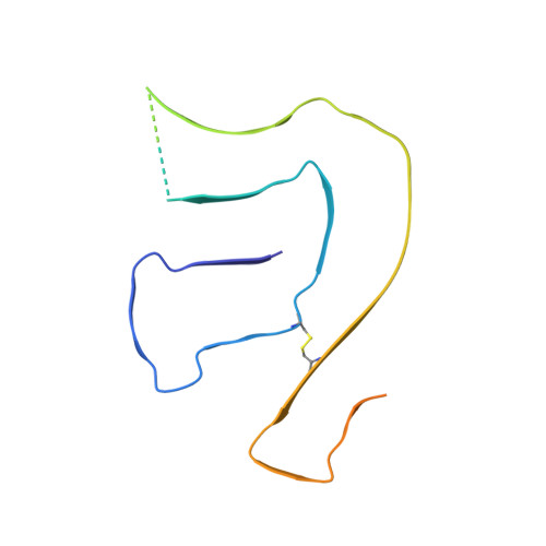
Cryo-EM structure of cardiac amyloid fibrils from an immunoglobulin light chain AL amyloidosis patient.

Swuec, P.Lavatelli, F.Tasaki, M.Paissoni, C.Rognoni, P.Maritan, M.Brambilla, F.Milani, P.Mauri, P.Camilloni, C.Palladini, G.Merlini, G.Ricagno, S.Bolognesi, M.

(2019) Nat Commun 10: 1269-1269

  • DOI: https://doi.org/10.1038/s41467-019-09133-w
  • Primary Citation of Related Structures:  
    6HUD

  • PubMed Abstract: 

    Systemic light chain amyloidosis (AL)  is a life-threatening disease caused by aggregation and deposition of monoclonal immunoglobulin light chains (LC) in target organs. Severity of heart involvement is the most important factor determining prognosis. Here, we report the 4.0 Å resolution cryo-electron microscopy map and molecular model of amyloid fibrils extracted from the heart of an AL amyloidosis patient with severe amyloid cardiomyopathy. The helical fibrils are composed of a single protofilament, showing typical 4.9 Å stacking and cross-β architecture. Two distinct polypeptide stretches (total of 77 residues) from the LC variable domain (V l ) fit the fibril density. Despite V l high sequence variability, residues stabilizing the fibril core are conserved through different cardiotoxic V l , highlighting structural motifs that may be common to misfolding-prone LCs. Our data shed light on the architecture of LC amyloids, correlate amino acid sequences with fibril assembly, providing the grounds for development of innovative medicines.


  • Organizational Affiliation

    Dipartimento di Bioscienze, Università degli Studi di Milano, Via Celoria 26, 20133, Milano, Italy.


Macromolecules
Find similar proteins by:  (by identity cutoff)  |  3D Structure
Entity ID: 1
MoleculeChains Sequence LengthOrganismDetailsImage
Monoclonal immunoglobulin light chains (LC)
A, B, C, D, E
136Homo sapiensMutation(s): 0 
Entity Groups  
Sequence Clusters30% Identity50% Identity70% Identity90% Identity95% Identity100% Identity
Sequence Annotations
Expand
  • Reference Sequence
Experimental Data & Validation

Experimental Data

  • Method: ELECTRON MICROSCOPY
  • Resolution: 4.00 Å
  • Aggregation State: FILAMENT 
  • Reconstruction Method: HELICAL 
EM Software:
TaskSoftware PackageVersion
RECONSTRUCTIONRELION2.1beta1
MODEL REFINEMENTPHENIX1.13

Structure Validation

View Full Validation Report


Entry History & Funding Information

Deposition Data


Funding OrganizationLocationGrant Number
Italian Association for Cancer ResearchItaly9965
Fondazione CARIPLO--
Italian Ministry of Health--
Italian Medicines Agency--

Revision History  (Full details and data files)

  • Version 1.0: 2019-03-27
    Type: Initial release
  • Version 1.1: 2019-04-03
    Changes: Data collection, Database references