


The crystal structure of type II Dehydroquinase from Psychromonas ingrahamii 37
Lapthorn, A.J., Koyroytsaltis-McQuire, D., Roszak, A.W.To be published.
Experimental Data Snapshot
Starting Model: experimental
View more details
Entity ID: 1 | |||||
|---|---|---|---|---|---|
| Molecule | Chains | Sequence Length | Organism | Details | Image |
| 3-dehydroquinate dehydratase | 152 | Psychromonas ingrahamii 37 | Mutation(s): 0 Gene Names: aroQ, Ping_3121 EC: 4.2.1.10 | 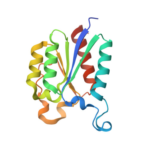 | |
UniProt | |||||
Entity Groups | |||||
| Sequence Clusters | 30% Identity50% Identity70% Identity90% Identity95% Identity100% Identity | ||||
| UniProt Group | A1SZA3 | ||||
Sequence AnnotationsExpand | |||||
Reference Sequence | |||||
| Ligands 3 Unique | |||||
|---|---|---|---|---|---|
| ID | Chains | Name / Formula / InChI Key | 2D Diagram | 3D Interactions | |
| MPO Download:Ideal Coordinates CCD File | Y [auth K] | 3[N-MORPHOLINO]PROPANE SULFONIC ACID C7 H15 N O4 S DVLFYONBTKHTER-UHFFFAOYSA-N |  | ||
| TLA Download:Ideal Coordinates CCD File | M [auth A] N [auth B] P [auth C] Q [auth D] R [auth E] | L(+)-TARTARIC ACID C4 H6 O6 FEWJPZIEWOKRBE-JCYAYHJZSA-N |  | ||
| GOL Download:Ideal Coordinates CCD File | O [auth B] | GLYCEROL C3 H8 O3 PEDCQBHIVMGVHV-UHFFFAOYSA-N |  | ||
| Length ( Å ) | Angle ( ˚ ) |
|---|---|
| a = 137.572 | α = 90 |
| b = 137.948 | β = 90 |
| c = 139.417 | γ = 90 |
| Software Name | Purpose |
|---|---|
| REFMAC | refinement |
| Aimless | data scaling |
| AMoRE | phasing |
| PDB_EXTRACT | data extraction |
| XDS | data reduction |
| Funding Organization | Location | Grant Number |
|---|---|---|
| Engineering and Physical Sciences Research Council | United Kingdom | EP/P00086X/1 |


