6HRC

Outward-facing PglK with ATPgammaS bound


Experimental Data Snapshot

  • Method: X-RAY DIFFRACTION
  • Resolution: 3.30 Å
  • R-Value Free: 0.284 
  • R-Value Work: 0.232 
  • R-Value Observed: 0.235 

wwPDB Validation   3D Report Full Report


Ligand Structure Quality Assessment 


This is version 1.2 of the entry. See complete history


Literature

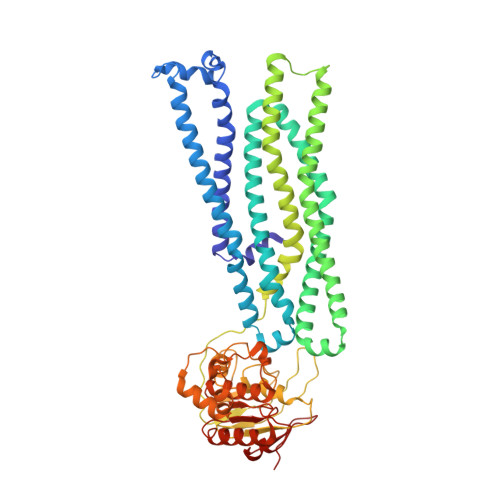
Structure of Outward-Facing PglK and Molecular Dynamics of Lipid-Linked Oligosaccharide Recognition and Translocation.

Perez, C.Mehdipour, A.R.Hummer, G.Locher, K.P.

(2019) Structure 27: 669-678.e5

  • DOI: https://doi.org/10.1016/j.str.2019.01.013
  • Primary Citation of Related Structures:  
    6HRC

  • PubMed Abstract: 

    PglK is a lipid-linked oligosaccharide (LLO) flippase essential for asparagine-linked protein glycosylation in Campylobacter jejuni. Previously we have proposed a non-alternating-access LLO translocation mechanism, where postulated outward-facing states play a primary role. To investigate this unusual mechanistic proposal, we have determined a high-resolution structure of PglK that displays an outward semi-occluded state with the two nucleotide binding domains forming an asymmetric closed dimer with two bound ATPγS molecules. Based on this structure, we performed extensive molecular dynamics simulations to investigate LLO recognition and flipping. Our results suggest that PglK may employ a "substrate-hunting" mechanism to locally increase the LLO concentration and facilitate its jump into the translocation pathway, for which sugars from the LLO head group are essential. We further conclude that the release of LLO to the outside occurs before ATP hydrolysis and is followed by the closing of the periplasmic cavity of PglK.


  • Organizational Affiliation

    Department of Biology, Institute of Molecular Biology and Biophysics, ETH Zürich, 8093 Zürich, Switzerland; Biozentrum, University of Basel, 4056 Basel, Switzerland.


Macromolecules
Find similar proteins by:  (by identity cutoff)  |  3D Structure
Entity ID: 1
MoleculeChains Sequence LengthOrganismDetailsImage
WlaB protein
A, B, C, D
564Campylobacter jejuniMutation(s): 1 
Gene Names: wlaB
Membrane Entity: Yes 
UniProt
Find proteins for O86150 (Campylobacter jejuni)
Explore O86150 
Go to UniProtKB:  O86150
Entity Groups  
Sequence Clusters30% Identity50% Identity70% Identity90% Identity95% Identity100% Identity
UniProt GroupO86150
Sequence Annotations
Expand
  • Reference Sequence
Experimental Data & Validation

Experimental Data

  • Method: X-RAY DIFFRACTION
  • Resolution: 3.30 Å
  • R-Value Free: 0.284 
  • R-Value Work: 0.232 
  • R-Value Observed: 0.235 
  • Space Group: P 21 21 21
Unit Cell:
Length ( Å )Angle ( ˚ )
a = 125.68α = 90
b = 145.98β = 90
c = 206.76γ = 90
Software Package:
Software NamePurpose
PHENIXrefinement
XDSdata reduction
XSCALEdata scaling
PHASERphasing
PDB_EXTRACTdata extraction

Structure Validation

View Full Validation Report


Ligand Structure Quality Assessment 


Entry History & Funding Information

Deposition Data


Funding OrganizationLocationGrant Number
Swiss National Science FoundationSwitzerland310030B_166672

Revision History  (Full details and data files)

  • Version 1.0: 2019-03-06
    Type: Initial release
  • Version 1.1: 2019-04-10
    Changes: Data collection, Database references
  • Version 1.2: 2024-01-24
    Changes: Data collection, Database references, Derived calculations, Refinement description