


Nitrocefin acylation of single catalytic serine of penicillin-binding protein 3 from P. aeruginosa
Bellini, D., Dowson, C.G.To be published.
Experimental Data Snapshot
Starting Model: experimental
View more details
Entity ID: 1 | |||||
|---|---|---|---|---|---|
| Molecule | Chains | Sequence Length | Organism | Details | Image |
| Peptidoglycan D,D-transpeptidase FtsI | 535 | Pseudomonas aeruginosa PAO1 | Mutation(s): 0 Gene Names: ftsI, pbpB, PA4418 EC: 3.4.16.4 | 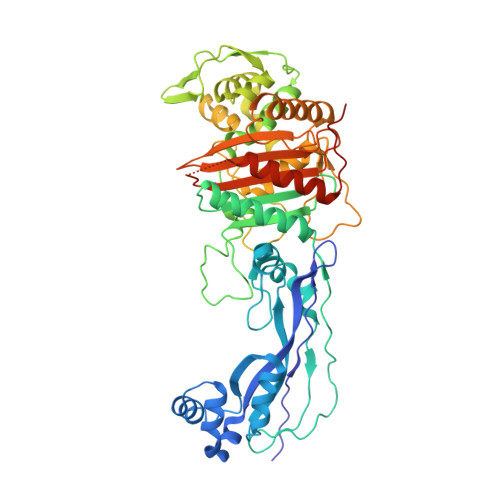 | |
UniProt | |||||
Entity Groups | |||||
| Sequence Clusters | 30% Identity50% Identity70% Identity90% Identity95% Identity100% Identity | ||||
| UniProt Group | G3XD46 | ||||
Sequence AnnotationsExpand | |||||
Reference Sequence | |||||
| Ligands 1 Unique | |||||
|---|---|---|---|---|---|
| ID | Chains | Name / Formula / InChI Key | 2D Diagram | 3D Interactions | |
| NEF (Subject of Investigation/LOI) Download:Ideal Coordinates CCD File | B [auth A] | Nitrocefin - open form C21 H18 N4 O9 S2 PYBZXZZHQKFSGC-CZHQAMEJSA-N |  | ||
| Length ( Å ) | Angle ( ˚ ) |
|---|---|
| a = 68.68 | α = 90 |
| b = 83.272 | β = 90 |
| c = 89.102 | γ = 90 |
| Software Name | Purpose |
|---|---|
| PHENIX | refinement |
| DIALS | data reduction |
| Aimless | data scaling |
| MOLREP | phasing |
| Funding Organization | Location | Grant Number |
|---|---|---|
| Medical Research Council (MRC, United Kingdom) | United Kingdom | grant.MR/P007503/1 |


