6HM6 | pdb_00006hm6

CRYSTAL STRUCTURE OF SPLEEN TYROSINE KINASE (SYK) IN COMPLEX WITH A 2-(PYRIDINYLOXYMETHYL)PYRIDINE INHIBITOR


Experimental Data Snapshot

  • Method: X-RAY DIFFRACTION
  • Resolution: 2.10 Å
  • R-Value Free: 
    0.290 (Depositor), 0.298 (DCC) 
  • R-Value Work: 
    0.213 (Depositor) 
  • R-Value Observed: 
    0.217 (Depositor) 

Starting Model: other
View more details

wwPDB Validation 3D Report Full Report

Validation slider image for 6HM6

Ligand Structure Quality Assessment 


This is version 1.3 of the entry. See complete history

Literature

Discovery of potent and selective Spleen Tyrosine Kinase inhibitors for the topical treatment of inflammatory skin disease.

Barker, M.D.Liddle, J.Atkinson, F.L.Wilson, D.M.Dickson, M.C.Ramirez-Molina, C.Lewis, H.Davis, R.P.Somers, D.O.Neu, M.Jones, E.Watson, R.

(2018) Bioorg Med Chem Lett 28: 3458-3462

  • DOI: https://doi.org/10.1016/j.bmcl.2018.09.022
  • Primary Citation Related Structures: 
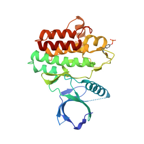
    6HM6, 6HM7

  • PubMed Abstract: 

    The discovery and lead optimisation of a novel series of SYK inhibitors is described. These were optimised for SYK potency and selectivity against Aurora B. Compounds were profiled in a human skin penetration study to identify a suitable candidate molecule for pre-clinical development. Compound 44 (GSK2646264) was selected for progression and is currently in Phase I clinical trials.


  • Organizational Affiliation
    • GlaxoSmithKline R&D, Medicines Research Centre, Gunnels Wood Road, Stevenage, Hertfordshire SG1 2NY, UK. Electronic address: mike.d.barker@gsk.com.

Macromolecule Content 

  • Total Structure Weight: 32.47 kDa 
  • Atom Count: 2,276 
  • Modeled Residue Count: 261 
  • Deposited Residue Count: 276 
  • Unique protein chains: 1

Macromolecules

Find similar proteins by:|  3D Structure
Entity ID: 1
MoleculeChains  Sequence LengthOrganismDetailsImage
SPLEEN TYROSINE KINASE276Homo sapiensMutation(s): 0 
EC: 2.7.10.2
UniProt & NIH Common Fund Data Resources
Find proteins for P43405 (Homo sapiens)
Explore P43405 
Go to UniProtKB:  P43405
PHAROS:  P43405
GTEx:  ENSG00000165025 
Entity Groups
Sequence Clusters30% Identity50% Identity70% Identity90% Identity95% Identity100% Identity
UniProt GroupP43405
Sequence Annotations
Expand
Reference Sequence

Small Molecules

Ligands 2 Unique
IDChains Name / Formula / InChI Key2D Diagram3D Interactions
GDK

Query on GDK


Download:Ideal Coordinates CCD File
B [auth A]2-(4-methylphenyl)-3-(pyridin-2-ylmethoxy)pyridine
C18 H16 N2 O
GEICNUUKZLWKET-UHFFFAOYSA-N
GOL

Query on GOL


Download:Ideal Coordinates CCD File
C [auth A]GLYCEROL
C3 H8 O3
PEDCQBHIVMGVHV-UHFFFAOYSA-N
Modified Residues  1 Unique
IDChains TypeFormula2D DiagramParent
PTR
Query on PTR
A
L-PEPTIDE LINKINGC9 H12 N O6 PTYR

Experimental Data & Validation

Experimental Data

  • Method: X-RAY DIFFRACTION
  • Resolution: 2.10 Å
  • R-Value Free:  0.290 (Depositor), 0.298 (DCC) 
  • R-Value Work:  0.213 (Depositor) 
  • R-Value Observed: 0.217 (Depositor) 
Space Group: P 1 21 1
Unit Cell:
Length ( Å )Angle ( ˚ )
a = 39.437α = 90
b = 84.982β = 100.5
c = 40.277γ = 90
Software Package:
Software NamePurpose
d*TREKdata reduction
d*TREKdata scaling
PDB_EXTRACTdata extraction
REFMACphasing
REFMACrefinement
Cootmodel building

Structure Validation

View Full Validation Report


Ligand Structure Quality Assessment 


Entry History 

Deposition Data

  • Released Date: 2018-10-10 
  • Deposition Author(s): Somers, D.O.

Revision History  (Full details and data files)

  • Version 1.0: 2018-10-10
    Type: Initial release
  • Version 1.1: 2018-10-17
    Changes: Data collection, Database references
  • Version 1.2: 2024-05-01
    Changes: Data collection, Database references, Refinement description
  • Version 1.3: 2024-11-06
    Changes: Structure summary