6HL8 | pdb_00006hl8

Crystal Structure of the CsiD Glutarate Hydroxylase in complex with Glutarate


Experimental Data Snapshot

  • Method: X-RAY DIFFRACTION
  • Resolution: 2.40 Å
  • R-Value Free: 
    0.220 (Depositor), 0.186 (DCC) 
  • R-Value Work: 
    0.175 (Depositor) 
  • R-Value Observed: 
    0.177 (Depositor) 

wwPDB Validation 3D Report Full Report

Validation slider image for 6HL8

This is version 1.2 of the entry. See complete history

Literature

Widespread bacterial lysine degradation proceeding via glutarate and L-2-hydroxyglutarate.

Knorr, S.Sinn, M.Galetskiy, D.Williams, R.M.Wang, C.Muller, N.Mayans, O.Schleheck, D.Hartig, J.S.

(2018) Nat Commun 9: 5071-5071

  • DOI: https://doi.org/10.1038/s41467-018-07563-6
  • Primary Citation Related Structures: 
    6GPE, 6GPN, 6HL8, 6HL9

  • PubMed Abstract: 

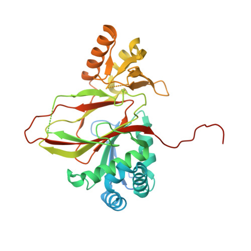
    Lysine degradation has remained elusive in many organisms including Escherichia coli. Here we report catabolism of lysine to succinate in E. coli involving glutarate and L-2-hydroxyglutarate as intermediates. We show that CsiD acts as an α-ketoglutarate-dependent dioxygenase catalysing hydroxylation of glutarate to L-2-hydroxyglutarate. CsiD is found widespread in bacteria. We present crystal structures of CsiD in complex with glutarate, succinate, and the inhibitor N-oxalyl-glycine, demonstrating strong discrimination between the structurally related ligands. We show that L-2-hydroxyglutarate is converted to α-ketoglutarate by LhgO acting as a membrane-bound, ubiquinone-linked dehydrogenase. Lysine enters the pathway via 5-aminovalerate by the promiscuous enzymes GabT and GabD. We demonstrate that repression of the pathway by CsiR is relieved upon glutarate binding. In conclusion, lysine degradation provides an important link in central metabolism. Our results imply the gut microbiome as a potential source of glutarate and L-2-hydroxyglutarate associated with human diseases such as cancer and organic acidurias.


  • Organizational Affiliation
    • Department of Chemistry, University of Konstanz, Konstanz, 78457, Germany.

Macromolecule Content 

  • Total Structure Weight: 81.67 kDa 
  • Atom Count: 5,111 
  • Modeled Residue Count: 583 
  • Deposited Residue Count: 706 
  • Unique protein chains: 1

Macromolecules

Find similar proteins by:|  3D Structure
Entity ID: 1
MoleculeChains  Sequence LengthOrganismDetailsImage
Protein CsiD
A, B
353Escherichia coli K-12Mutation(s): 0 
Gene Names: csiDgabygaTb2659JW5427
EC: 1.14.11.64
UniProt
Find proteins for P76621 (Escherichia coli (strain K12))
Explore P76621 
Go to UniProtKB:  P76621
Entity Groups
Sequence Clusters30% Identity50% Identity70% Identity90% Identity95% Identity100% Identity
UniProt GroupP76621
Sequence Annotations
Expand
Reference Sequence

Experimental Data & Validation

Experimental Data

  • Method: X-RAY DIFFRACTION
  • Resolution: 2.40 Å
  • R-Value Free:  0.220 (Depositor), 0.186 (DCC) 
  • R-Value Work:  0.175 (Depositor) 
  • R-Value Observed: 0.177 (Depositor) 
Space Group: P 4 21 2
Unit Cell:
Length ( Å )Angle ( ˚ )
a = 120.8α = 90
b = 120.8β = 90
c = 136.43γ = 90
Software Package:
Software NamePurpose
PHENIXrefinement
XDSdata reduction
XSCALEdata scaling
PHASERphasing
PDB_EXTRACTdata extraction

Structure Validation

View Full Validation Report


Entry History 

Deposition Data

Revision History  (Full details and data files)

  • Version 1.0: 2018-12-12
    Type: Initial release
  • Version 1.1: 2023-02-08
    Changes: Advisory, Database references, Derived calculations
  • Version 1.2: 2024-06-19
    Changes: Data collection