6H96 | pdb_00006h96

AlbA-albicidin complex, albicidin resistance protein


Experimental Data Snapshot

  • Method: X-RAY DIFFRACTION
  • Resolution: 1.55 Å
  • R-Value Free: 
    0.207 (Depositor), 0.215 (DCC) 
  • R-Value Work: 
    0.189 (Depositor), 0.197 (DCC) 
  • R-Value Observed: 
    0.190 (Depositor) 

Starting Model: experimental
View more details

wwPDB Validation 3D Report Full Report

Validation slider image for 6H96

Ligand Structure Quality Assessment 


This is version 1.3 of the entry. See complete history

Literature

Adaptation of a Bacterial Multidrug Resistance System Revealed by the Structure and Function of AlbA.

Sikandar, A.Cirnski, K.Testolin, G.Volz, C.Bronstrup, M.Kalinina, O.V.Muller, R.Koehnke, J.

(2018) J Am Chem Soc 140: 16641-16649

  • DOI: https://doi.org/10.1021/jacs.8b08895
  • Primary Citation Related Structures: 
    6H95, 6H96, 6H97, 6HAI

  • PubMed Abstract: 

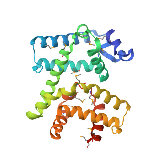
    To combat the rise of antimicrobial resistance, the discovery of new antibiotics is paramount. Albicidin and cystobactamid are related natural product antibiotics with potent activity against Gram-positive and, crucially, Gram-negative pathogens. AlbA has been reported to neutralize albicidin by binding it with nanomolar affinity. To understand this potential resistance mechanism, we determined structures of AlbA and its complex with albicidin. The structures revealed AlbA to be comprised of two domains, each unexpectedly resembling the multiantibiotic neutralizing protein TipA. Binding of the long albicidin molecule was shared pseudosymmetrically between the two domains. The structure also revealed an unexpected chemical modification of albicidin, which we demonstrate to be promoted by AlbA, and to reduce albicidin potency; we propose a mechanism for this reaction. Overall, our findings suggest that AlbA arose through internal duplication in an ancient TipA-like gene, leading to a new binding scaffold adapted to the sequestration of long-chain antibiotics.


  • Organizational Affiliation
    • Workgroup Structural Biology of Biosynthetic Enzymes, Helmholtz Institute for Pharmaceutical Research Saarland , Helmholtz Centre for Infection Research, Saarland University , Campus Geb. E8.1 , Saarbrücken 66123 , Germany.

Macromolecule Content 

  • Total Structure Weight: 56.06 kDa 
  • Atom Count: 4,146 
  • Modeled Residue Count: 429 
  • Deposited Residue Count: 452 
  • Unique protein chains: 1

Macromolecules

Find similar proteins by:|  3D Structure
Entity ID: 1
MoleculeChains  Sequence LengthOrganismDetailsImage
Albicidin resistance protein
A, B
226Klebsiella oxytocaMutation(s): 0 
Gene Names: albA
UniProt
Find proteins for Q8KRS7 (Klebsiella oxytoca)
Explore Q8KRS7 
Go to UniProtKB:  Q8KRS7
Entity Groups
Sequence Clusters30% Identity50% Identity70% Identity90% Identity95% Identity100% Identity
UniProt GroupQ8KRS7
Sequence Annotations
Expand
Reference Sequence

Small Molecules

Ligands 2 Unique
IDChains Name / Formula / InChI Key2D Diagram3D Interactions
FWE

Query on FWE


Download:Ideal Coordinates CCD File
C [auth A],
E [auth B]
4-[[4-[[4-[(3~{S})-5-azanyl-3-[[4-[[(~{E})-3-(4-hydroxyphenyl)-2-methyl-prop-2-enoyl]amino]phenyl]carbonylamino]-2-oxidanylidene-3~{H}-pyrrol-1-yl]phenyl]carbonylamino]-3-methoxy-2-oxidanyl-phenyl]carbonylamino]-3-methoxy-2-oxidanyl-benzoic acid
C44 H38 N6 O12
VHKFIJVTNPVMBS-QCKJEFJWSA-N
SO4

Query on SO4


Download:Ideal Coordinates CCD File
D [auth A],
F [auth B]
SULFATE ION
O4 S
QAOWNCQODCNURD-UHFFFAOYSA-L
Modified Residues  1 Unique
IDChains TypeFormula2D DiagramParent
MSE
Query on MSE
A, B
L-PEPTIDE LINKINGC5 H11 N O2 SeMET

Experimental Data & Validation

Experimental Data

  • Method: X-RAY DIFFRACTION
  • Resolution: 1.55 Å
  • R-Value Free:  0.207 (Depositor), 0.215 (DCC) 
  • R-Value Work:  0.189 (Depositor), 0.197 (DCC) 
  • R-Value Observed: 0.190 (Depositor) 
Space Group: C 2 2 21
Unit Cell:
Length ( Å )Angle ( ˚ )
a = 54.142α = 90
b = 123.181β = 90
c = 161.06γ = 90
Software Package:
Software NamePurpose
PHENIXrefinement
xia2data reduction
xia2data scaling
PHASERphasing

Structure Validation

View Full Validation Report


Ligand Structure Quality Assessment 


Entry History 

& Funding Information

Deposition Data


Funding OrganizationLocationGrant Number
GermanyKO4116_3_1

Revision History  (Full details and data files)

  • Version 1.0: 2018-11-21
    Type: Initial release
  • Version 1.1: 2019-04-24
    Changes: Data collection, Database references
  • Version 1.2: 2024-05-01
    Changes: Data collection, Database references, Refinement description
  • Version 1.3: 2024-10-23
    Changes: Structure summary