6H7X | pdb_00006h7x

First X-ray structure of full-length human RuvB-Like 2.


Experimental Data Snapshot

  • Method: X-RAY DIFFRACTION
  • Resolution: 2.89 Å
  • R-Value Free: 
    0.237 (Depositor), 0.237 (DCC) 
  • R-Value Work: 
    0.205 (Depositor), 0.202 (DCC) 
  • R-Value Observed: 
    0.206 (Depositor) 

Starting Model: experimental
View more details

wwPDB Validation 3D Report Full Report

Validation slider image for 6H7X

This is version 1.3 of the entry. See complete history

Literature

X-ray structure of full-length human RuvB-Like 2 - mechanistic insights into coupling between ATP binding and mechanical action.

Silva, S.T.N.Brito, J.A.Arranz, R.Sorzano, C.O.S.Ebel, C.Doutch, J.Tully, M.D.Carazo, J.M.Carrascosa, J.L.Matias, P.M.Bandeiras, T.M.

(2018) Sci Rep 8: 13726-13726

  • DOI: https://doi.org/10.1038/s41598-018-31997-z
  • Primary Citation Related Structures: 
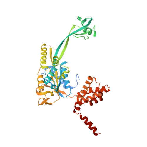
    6H7X

  • PubMed Abstract: 

    RuvB-Like transcription factors function in cell cycle regulation, development and human disease, such as cancer and heart hyperplasia. The mechanisms that regulate adenosine triphosphate (ATP)-dependent activity, oligomerization and post-translational modifications in this family of enzymes are yet unknown. We present the first crystallographic structure of full-length human RuvBL2 which provides novel insights into its mechanistic action and biology. The ring-shaped hexameric RuvBL2 structure presented here resolves for the first time the mobile domain II of the human protein, which is responsible for protein-protein interactions and ATPase activity regulation. Structural analysis suggests how ATP binding may lead to domain II motion through interactions with conserved N-terminal loop histidine residues. Furthermore, a comparison between hsRuvBL1 and 2 shows differences in surface charge distribution that may account for previously described differences in regulation. Analytical ultracentrifugation and cryo electron microscopy analyses performed on hsRuvBL2 highlight an oligomer plasticity that possibly reflects different physiological conformations of the protein in the cell, as well as that single-stranded DNA (ssDNA) can promote the oligomerization of monomeric hsRuvBL2. Based on these findings, we propose a mechanism for ATP binding and domain II conformational change coupling.


  • Organizational Affiliation
    • Instituto de Tecnologia Química e Biológica António Xavier, Universidade Nova de Lisboa, Av. da República, 2780-157, Oeiras, Portugal.

Macromolecule Content 

  • Total Structure Weight: 51.84 kDa 
  • Atom Count: 2,962 
  • Modeled Residue Count: 371 
  • Deposited Residue Count: 463 
  • Unique protein chains: 1

Macromolecules

Find similar proteins by:|  3D Structure
Entity ID: 1
MoleculeChains  Sequence LengthOrganismDetailsImage
RuvB-like 2463Homo sapiensMutation(s): 0 
Gene Names: RUVBL2INO80JTIP48TIP49BCGI-46
EC: 3.6.4.12
UniProt & NIH Common Fund Data Resources
Find proteins for Q9Y230 (Homo sapiens)
Explore Q9Y230 
Go to UniProtKB:  Q9Y230
PHAROS:  Q9Y230
GTEx:  ENSG00000183207 
Entity Groups
Sequence Clusters30% Identity50% Identity70% Identity90% Identity95% Identity100% Identity
UniProt GroupQ9Y230
Sequence Annotations
Expand
Reference Sequence

Small Molecules

Ligands 3 Unique
IDChains Name / Formula / InChI Key2D Diagram3D Interactions
PEG

Query on PEG


Download:Ideal Coordinates CCD File
F [auth A]DI(HYDROXYETHYL)ETHER
C4 H10 O3
MTHSVFCYNBDYFN-UHFFFAOYSA-N
EDO

Query on EDO


Download:Ideal Coordinates CCD File
E [auth A]
G [auth A]
H [auth A]
I [auth A]
J [auth A]
E [auth A],
G [auth A],
H [auth A],
I [auth A],
J [auth A],
K [auth A],
L [auth A]
1,2-ETHANEDIOL
C2 H6 O2
LYCAIKOWRPUZTN-UHFFFAOYSA-N
MG

Query on MG


Download:Ideal Coordinates CCD File
B [auth A],
C [auth A],
D [auth A]
MAGNESIUM ION
Mg
JLVVSXFLKOJNIY-UHFFFAOYSA-N

Experimental Data & Validation

Experimental Data

  • Method: X-RAY DIFFRACTION
  • Resolution: 2.89 Å
  • R-Value Free:  0.237 (Depositor), 0.237 (DCC) 
  • R-Value Work:  0.205 (Depositor), 0.202 (DCC) 
  • R-Value Observed: 0.206 (Depositor) 
Space Group: P 6
Unit Cell:
Length ( Å )Angle ( ˚ )
a = 123.023α = 90
b = 123.023β = 90
c = 60.88γ = 120
Software Package:
Software NamePurpose
PHENIXrefinement
autoPROCdata collection
BUCCANEERmodel building
PHASERphasing
XDSdata reduction
Aimlessdata scaling

Structure Validation

View Full Validation Report


Entry History 

& Funding Information

Deposition Data


Funding OrganizationLocationGrant Number
Other governmentPortugalSFRH/BD/78706/2011/

Revision History  (Full details and data files)

  • Version 1.0: 2018-08-08
    Type: Initial release
  • Version 1.1: 2018-09-19
    Changes: Data collection, Database references
  • Version 1.2: 2018-09-26
    Changes: Data collection, Database references
  • Version 1.3: 2024-01-17
    Changes: Data collection, Database references, Refinement description, Structure summary