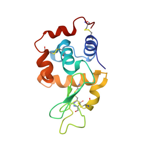
6H3B

Lysozyme: Machining protein microcrystals for structure determination by electron diffraction

  • Classification: HYDROLASE
  • Organism(s): Gallus gallus
  • Mutation(s): No 

  • Deposited: 2018-07-18 Released: 2018-09-12 
  • Deposition Author(s): Duyvesteyn, H.M.E., Ginn, H.M., Stuart, D.I.
  • Funding Organization(s): Medical Research Council (United Kingdom), Wellcome Trust, National Institutes of Health/National Human Genome Research Institute (NIH/NHGRI)

Experimental Data Snapshot

  • Method: ELECTRON CRYSTALLOGRAPHY
  • Resolution: 1.90 Å
  • R-Value Free: 0.283 
  • R-Value Work: 0.291 

wwPDB Validation   3D Report Full Report


This is version 1.3 of the entry. See complete history


Literature

Machining protein microcrystals for structure determination by electron diffraction.

Duyvesteyn, H.M.E.Kotecha, A.Ginn, H.M.Hecksel, C.W.Beale, E.V.de Haas, F.Evans, G.Zhang, P.Chiu, W.Stuart, D.I.

(2018) Proc Natl Acad Sci U S A 115: 9569-9573

  • DOI: https://doi.org/10.1073/pnas.1809978115
  • Primary Citation of Related Structures:  
    6H3B

  • PubMed Abstract: 

    We demonstrate that ion-beam milling of frozen, hydrated protein crystals to thin lamella preserves the crystal lattice to near-atomic resolution. This provides a vehicle for protein structure determination, bridging the crystal size gap between the nanometer scale of conventional electron diffraction and micron scale of synchrotron microfocus beamlines. The demonstration that atomic information can be retained suggests that milling could provide such detail on sections cut from vitrified cells.


  • Organizational Affiliation

    Division of Structural Biology, University of Oxford, Headington, Oxford OX3 7BN, United Kingdom.


Macromolecules
Find similar proteins by:  (by identity cutoff)  |  3D Structure
Entity ID: 1
MoleculeChains Sequence LengthOrganismDetailsImage
Lysozyme C129Gallus gallusMutation(s): 0 
EC: 3.2.1.17
UniProt
Find proteins for P00698 (Gallus gallus)
Explore P00698 
Go to UniProtKB:  P00698
Entity Groups  
Sequence Clusters30% Identity50% Identity70% Identity90% Identity95% Identity100% Identity
UniProt GroupP00698
Sequence Annotations
Expand
  • Reference Sequence
Small Molecules
Ligands 1 Unique
IDChains Name / Formula / InChI Key2D Diagram3D Interactions
CL
Query on CL

Download Ideal Coordinates CCD File 
B [auth A]CHLORIDE ION
Cl
VEXZGXHMUGYJMC-UHFFFAOYSA-M
Experimental Data & Validation

Experimental Data

  • Method: ELECTRON CRYSTALLOGRAPHY
  • Resolution: 1.90 Å
  • R-Value Free: 0.283 
  • R-Value Work: 0.291 
  • Space Group: P 43 21 2
Unit Cell:
Length ( Å )Angle ( ˚ )
a = 79.9α = 90
b = 79.9β = 90
c = 38.67γ = 90

Structure Validation

View Full Validation Report


Entry History & Funding Information

Deposition Data


Funding OrganizationLocationGrant Number
Medical Research Council (United Kingdom)United KingdomMR/N00065X/1
Wellcome TrustUnited KingdomALR00750-B500.1
Wellcome TrustUnited KingdomALR00040
Wellcome TrustUnited Kingdom206422/Z/17/Z
Wellcome TrustUnited Kingdom101122/Z/13/Z
National Institutes of Health/National Human Genome Research Institute (NIH/NHGRI)United StatesP41GM103832
Wellcome TrustUnited Kingdom090532/Z/09/Z

Revision History  (Full details and data files)

  • Version 1.0: 2018-09-12
    Type: Initial release
  • Version 1.1: 2018-09-26
    Changes: Data collection, Database references
  • Version 1.2: 2022-03-30
    Changes: Author supporting evidence, Database references
  • Version 1.3: 2024-11-13
    Changes: Data collection, Structure summary