6H2K | pdb_00006h2k

Structure of BlaC from Mycobacterium tuberculosis bound to the trans-enamine adduct of sulbactam.


Experimental Data Snapshot

  • Method: X-RAY DIFFRACTION
  • Resolution: 1.90 Å
  • R-Value Free: 
    0.227 (Depositor), 0.234 (DCC) 
  • R-Value Work: 
    0.182 (Depositor), 0.192 (DCC) 
  • R-Value Observed: 
    0.184 (Depositor) 

Starting Model: experimental
View more details

wwPDB Validation 3D Report Full Report

Validation slider image for 6H2K

Ligand Structure Quality Assessment 


This is version 1.3 of the entry. See complete history

Literature

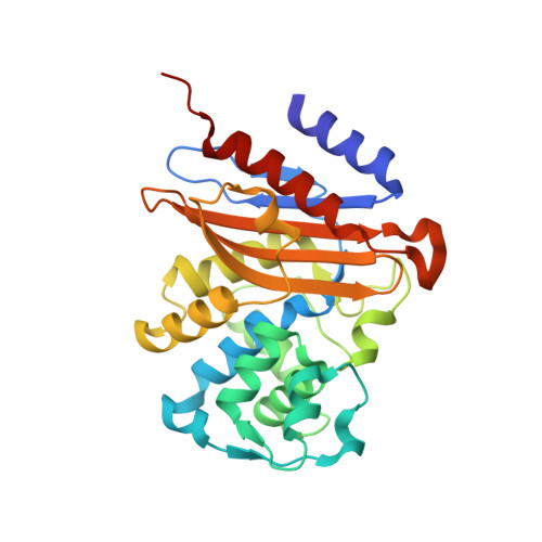
New Conformations of Acylation Adducts of Inhibitors of beta-Lactamase from Mycobacterium tuberculosis.

Tassoni, R.Blok, A.Pannu, N.S.Ubbink, M.

(2019) Biochemistry 58: 997-1009

  • DOI: https://doi.org/10.1021/acs.biochem.8b01085
  • Primary Citation Related Structures: 
    6H27, 6H28, 6H2A, 6H2C, 6H2G, 6H2H, 6H2I, 6H2K

  • PubMed Abstract: 

    Mycobacterium tuberculosis (Mtb), the main causative agent of tuberculosis (TB), is naturally resistant to β-lactam antibiotics due to the production of the extended spectrum β-lactamase BlaC. β-Lactam/β-lactamase inhibitor combination therapies can circumvent the BlaC-mediated resistance of Mtb and are promising treatment options against TB. However, still little is known of the exact mechanism of BlaC inhibition by the β-lactamase inhibitors currently approved for clinical use, clavulanic acid, sulbactam, tazobactam, and avibactam. Here, we present the X-ray diffraction crystal structures of the acyl-enzyme adducts of wild-type BlaC with the four inhibitors. The +70 Da adduct derived from clavulanate and the trans-enamine acylation adducts of sulbactam and tazobactam are reported. BlaC in complex with avibactam revealed two inhibitor conformations. Preacylation binding could not be observed because inhibitor binding was not detected in BlaC variants carrying a substitution of the active site serine 70 to either alanine or cysteine, by crystallography, ITC or NMR. These results suggest that the catalytic serine 70 is necessary not only for enzyme acylation but also for increasing BlaC affinity for inhibitors in the preacylation state. The structure of BlaC with the serine to cysteine mutation showed a covalent linkage of the cysteine 70 Sγ atom to the nearby amino group of lysine 73. The differences of adduct conformations between BlaC and other β-lactamases are discussed.


  • Organizational Affiliation
    • Leiden Institute of Chemistry , Leiden University , Einsteinweg 55 , Leiden 2333CC , The Netherlands.

Macromolecule Content 

  • Total Structure Weight: 60.63 kDa 
  • Atom Count: 4,490 
  • Modeled Residue Count: 534 
  • Deposited Residue Count: 548 
  • Unique protein chains: 1

Macromolecules

Find similar proteins by:|  3D Structure
Entity ID: 1
MoleculeChains  Sequence LengthOrganismDetailsImage
Beta-lactamase
A, B
274Mycobacterium tuberculosisMutation(s): 0 
Gene Names: blaCERS027646_02769
EC: 3.5.2.6

Small Molecules

Ligands 4 Unique
IDChains Name / Formula / InChI Key2D Diagram3D Interactions
TSL

Query on TSL


Download:Ideal Coordinates CCD File
C [auth A],
N [auth B]
TRANS-ENAMINE INTERMEDIATE OF SULBACTAM
C8 H13 N O5 S
DPHUOYJKDIRKGT-YUDCMIJISA-N
PEG

Query on PEG


Download:Ideal Coordinates CCD File
D [auth A]
E [auth A]
F [auth A]
G [auth A]
H [auth A]
D [auth A],
E [auth A],
F [auth A],
G [auth A],
H [auth A],
O [auth B]
DI(HYDROXYETHYL)ETHER
C4 H10 O3
MTHSVFCYNBDYFN-UHFFFAOYSA-N
GOL

Query on GOL


Download:Ideal Coordinates CCD File
I [auth A],
J [auth A],
P [auth B]
GLYCEROL
C3 H8 O3
PEDCQBHIVMGVHV-UHFFFAOYSA-N
ACT

Query on ACT


Download:Ideal Coordinates CCD File
K [auth A],
L [auth A],
M [auth A],
Q [auth B],
R [auth B]
ACETATE ION
C2 H3 O2
QTBSBXVTEAMEQO-UHFFFAOYSA-M

Experimental Data & Validation

Experimental Data

  • Method: X-RAY DIFFRACTION
  • Resolution: 1.90 Å
  • R-Value Free:  0.227 (Depositor), 0.234 (DCC) 
  • R-Value Work:  0.182 (Depositor), 0.192 (DCC) 
  • R-Value Observed: 0.184 (Depositor) 
Space Group: P 1
Unit Cell:
Length ( Å )Angle ( ˚ )
a = 39.71α = 105.1
b = 41.95β = 89.95
c = 76.93γ = 90.81
Software Package:
Software NamePurpose
REFMACrefinement
XDSdata reduction
Aimlessdata scaling
MOLREPphasing

Structure Validation

View Full Validation Report


Ligand Structure Quality Assessment 


Entry History 

Deposition Data

Revision History  (Full details and data files)

  • Version 1.0: 2019-01-23
    Type: Initial release
  • Version 1.1: 2019-02-27
    Changes: Data collection, Database references
  • Version 1.2: 2024-01-17
    Changes: Data collection, Database references, Refinement description
  • Version 1.3: 2024-11-20
    Changes: Structure summary