6H0W | pdb_00006h0w

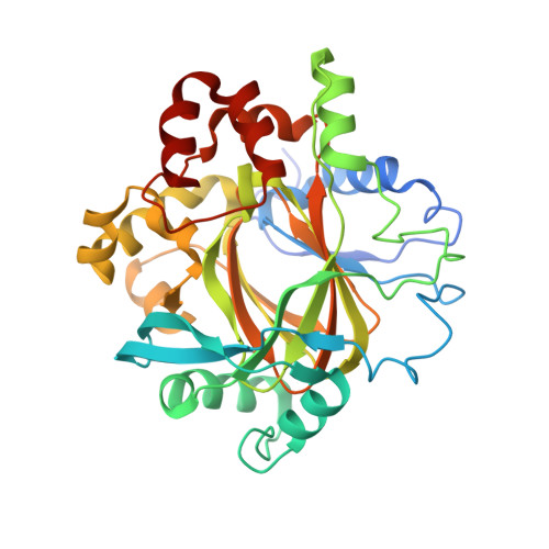
Crystal Structure of KDM4D with tetrazolylhydrazide ligand NS035


Experimental Data Snapshot

  • Method: X-RAY DIFFRACTION
  • Resolution: 1.23 Å
  • R-Value Free: 
    0.136 (Depositor), 0.143 (DCC) 
  • R-Value Work: 
    0.120 (Depositor), 0.134 (DCC) 
  • R-Value Observed: 
    0.120 (Depositor) 

Starting Model: experimental
View more details

wwPDB Validation 3D Report Full Report

Validation slider image for 6H0W

Ligand Structure Quality Assessment 


This is version 1.2 of the entry. See complete history

Literature

Structure-based mapping of the histone-binding pocket of KDM4D using functionalized tetrazole and pyridine core compounds.

Malecki, P.H.Fassauer, G.M.Ruger, N.Schulig, L.Link, A.Krylova, O.Heinemann, U.Weiss, M.S.

(2024) Eur J Med Chem 276: 116642-116642

  • DOI: https://doi.org/10.1016/j.ejmech.2024.116642
  • Primary Citation Related Structures: 
    6F5Q, 6F5R, 6F5S, 6F5T, 6H0W, 6H0X, 6H0Y, 6H0Z, 6H10, 6H11

  • PubMed Abstract: 

    KDM4 histone demethylases became an exciting target for inhibitor development as the evidence linking them directly to tumorigenesis mounts. In this study, we set out to better understand the binding cavity using an X-ray crystallographic approach to provide a detailed landscape of possible interactions within the under-investigated region of KDM4. Our design strategy was based on utilizing known KDM binding motifs, such as nicotinic acid and tetrazolylhydrazides, as core motifs that we decided to enrich with flexible tails to map the distal histone binding site. The resulting X-ray structures of the novel compounds bound to KDM4D, a representative of the KDM4 family, revealed the interaction pattern with distal residues in the histone-binding site. The most prominent protein rearrangement detected upon ligand binding is the loop movement that blocks the accessibility to the histone binding site. Apart from providing new sites that potential inhibitors can target, the novel compounds may prove helpful in exploring the capacity of ligands to bind in sites distal to the cofactor-binding site of other KDMs or 2-oxoglutarate (2OG)-dependent oxygenases. The case study proves that combining a strong small binding motif with flexible tails to probe the binding pocket will facilitate lead discovery in classical drug-discovery campaigns, given the ease of accessing X-ray quality crystals.


  • Organizational Affiliation
    • Macromolecular Structure and Interaction, Max-Delbrück-Center for Molecular Medicine, Robert-Rössle-Str. 10, 13125, Berlin, Germany; Macromolecular Crystallography, Helmholtz-Zentrum Berlin für Materialien und Energie, Albert-Einstein-Str. 15, 12489, Berlin, Germany; Department of Structural Biology of Prokaryotic Organisms, Institute of Bioorganic Chemistry, Polish Academy of Sciences, Z. Noskowskiego-Str. 12/14, 61-704, Poznań, Poland. Electronic address: pimalecki@ibch.poznan.pl.

Macromolecule Content 

  • Total Structure Weight: 40.94 kDa 
  • Atom Count: 3,547 
  • Modeled Residue Count: 330 
  • Deposited Residue Count: 342 
  • Unique protein chains: 1

Macromolecules

Find similar proteins by:|  3D Structure
Entity ID: 1
MoleculeChains  Sequence LengthOrganismDetailsImage
Lysine-specific demethylase 4D342Homo sapiensMutation(s): 0 
Gene Names: KDM4DJHDM3DJMJD2D
EC: 1.14.11 (PDB Primary Data), 1.14.11.66 (UniProt)
UniProt & NIH Common Fund Data Resources
Find proteins for Q6B0I6 (Homo sapiens)
Explore Q6B0I6 
Go to UniProtKB:  Q6B0I6
PHAROS:  Q6B0I6
GTEx:  ENSG00000186280 
Entity Groups
Sequence Clusters30% Identity50% Identity70% Identity90% Identity95% Identity100% Identity
UniProt GroupQ6B0I6
Sequence Annotations
Expand
Reference Sequence

Small Molecules

Ligands 9 Unique
IDChains Name / Formula / InChI Key2D Diagram3D Interactions
EPE

Query on EPE


Download:Ideal Coordinates CCD File
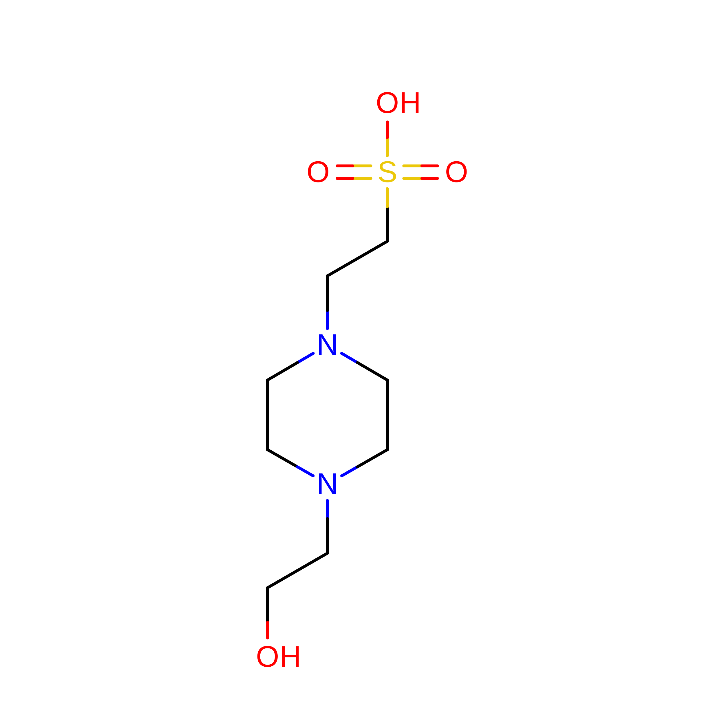
Q [auth A]4-(2-HYDROXYETHYL)-1-PIPERAZINE ETHANESULFONIC ACID
C8 H18 N2 O4 S
JKMHFZQWWAIEOD-UHFFFAOYSA-N
FHW

Query on FHW


Download:Ideal Coordinates CCD File
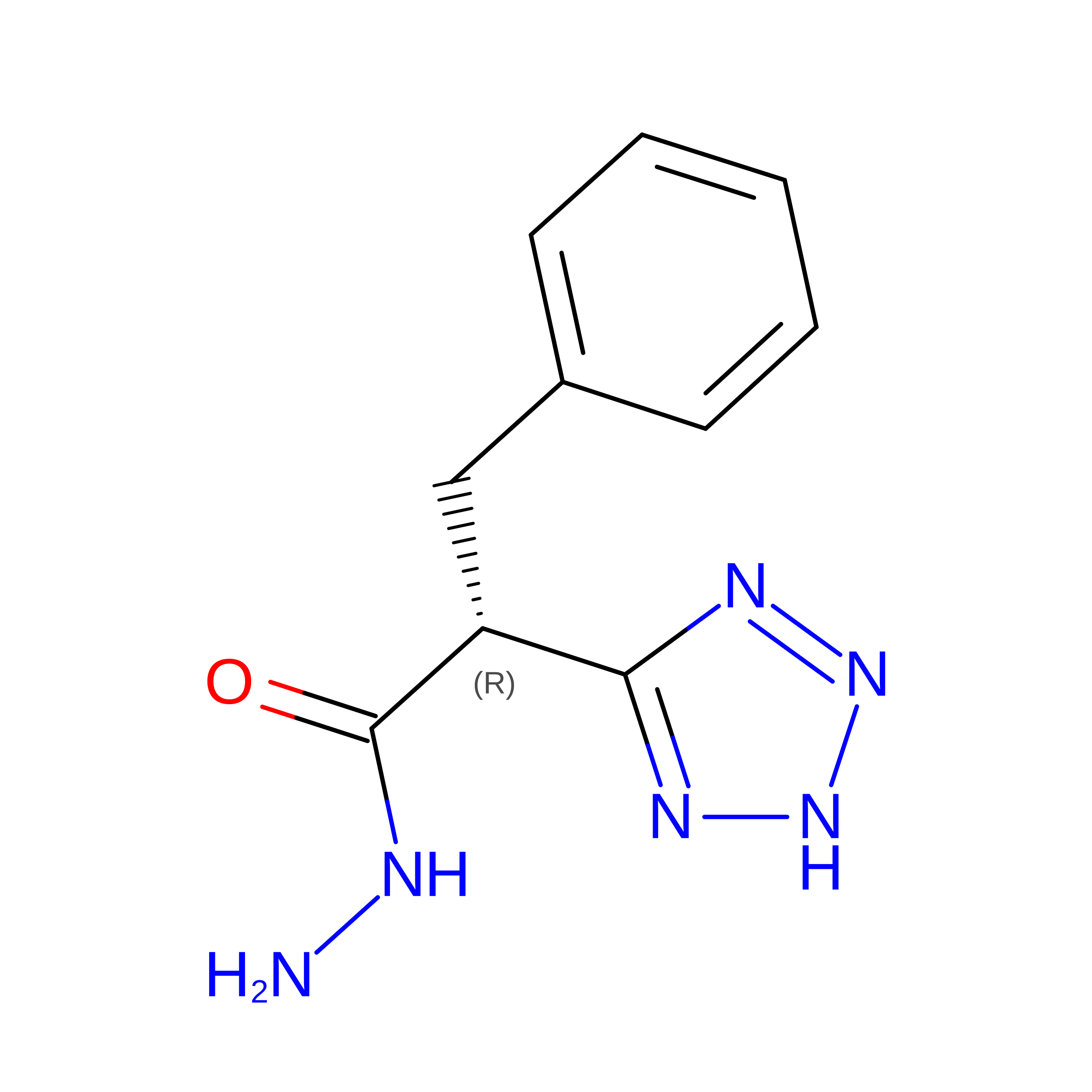
P [auth A](2~{R})-3-phenyl-2-(2~{H}-1,2,3,4-tetrazol-5-yl)propanehydrazide
C10 H12 N6 O
SGFUQCKYWZCXFN-MRVPVSSYSA-N
I1L

Query on I1L


Download:Ideal Coordinates CCD File
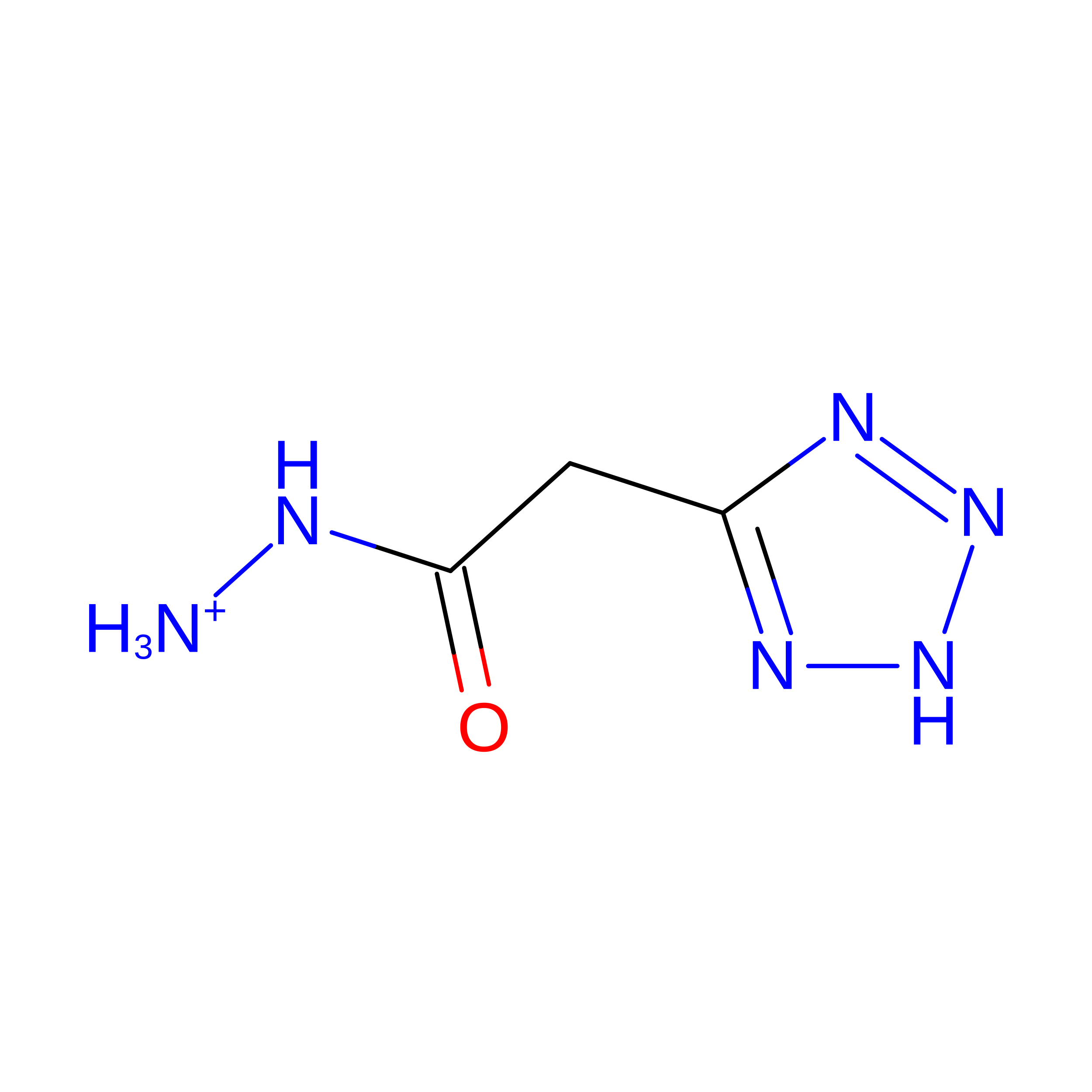
R [auth A][2-(2~{H}-1,2,3,4-tetrazol-5-yl)ethanoylamino]azanium
C3 H7 N6 O
TWKUAKLZOIABHJ-UHFFFAOYSA-O
SO4

Query on SO4


Download:Ideal Coordinates CCD File
M [auth A]SULFATE ION
O4 S
QAOWNCQODCNURD-UHFFFAOYSA-L
ZN

Query on ZN


Download:Ideal Coordinates CCD File
B [auth A]ZINC ION
Zn
PTFCDOFLOPIGGS-UHFFFAOYSA-N
EDO

Query on EDO


Download:Ideal Coordinates CCD File
D [auth A]
E [auth A]
F [auth A]
G [auth A]
H [auth A]
D [auth A],
E [auth A],
F [auth A],
G [auth A],
H [auth A],
I [auth A],
J [auth A],
K [auth A],
L [auth A]
1,2-ETHANEDIOL
C2 H6 O2
LYCAIKOWRPUZTN-UHFFFAOYSA-N
NI

Query on NI


Download:Ideal Coordinates CCD File
N [auth A]NICKEL (II) ION
Ni
VEQPNABPJHWNSG-UHFFFAOYSA-N
CL

Query on CL


Download:Ideal Coordinates CCD File
O [auth A]CHLORIDE ION
Cl
VEXZGXHMUGYJMC-UHFFFAOYSA-M
NA

Query on NA


Download:Ideal Coordinates CCD File
C [auth A]SODIUM ION
Na
FKNQFGJONOIPTF-UHFFFAOYSA-N

Experimental Data & Validation

Experimental Data

  • Method: X-RAY DIFFRACTION
  • Resolution: 1.23 Å
  • R-Value Free:  0.136 (Depositor), 0.143 (DCC) 
  • R-Value Work:  0.120 (Depositor), 0.134 (DCC) 
  • R-Value Observed: 0.120 (Depositor) 
Space Group: P 43 21 2
Unit Cell:
Length ( Å )Angle ( ˚ )
a = 71.482α = 90
b = 71.482β = 90
c = 150.707γ = 90
Software Package:
Software NamePurpose
XDSdata reduction
XSCALEdata scaling
PHENIXrefinement
PDB_EXTRACTdata extraction
REFMACphasing

Structure Validation

View Full Validation Report


Ligand Structure Quality Assessment 


Entry History 

Deposition Data

Revision History  (Full details and data files)

  • Version 1.0: 2020-01-29
    Type: Initial release
  • Version 1.1: 2024-01-17
    Changes: Data collection, Database references, Derived calculations, Refinement description
  • Version 1.2: 2026-07-22
    Changes: Database references, Structure summary