6GII | pdb_00006gii

The crystal structure of Tepidiphilus thermophilus P450 heme domain


Experimental Data Snapshot

  • Method: X-RAY DIFFRACTION
  • Resolution: 1.90 Å
  • R-Value Free: 
    0.228 (Depositor), 0.230 (DCC) 
  • R-Value Work: 
    0.178 (Depositor), 0.180 (DCC) 
  • R-Value Observed: 
    0.181 (Depositor) 

Starting Model: experimental
View more details

wwPDB Validation   3D Report Full Report


Ligand Structure Quality Assessment 

Created with Raphaël 2.3.0Worse 01 BetterLigand structure goodness of fit to experimental dataBest fitted HEMClick on this verticalbar to view details

This is version 1.2 of the entry. See complete history


Literature

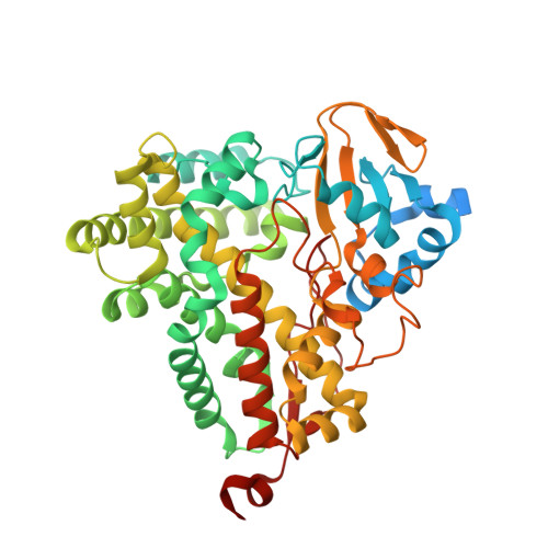
The crystal structure of P450-TT heme-domain provides the first structural insights into the versatile class VII P450s.

Tavanti, M.Porter, J.L.Levy, C.W.Gomez Castellanos, J.R.Flitsch, S.L.Turner, N.J.

(2018) Biochem Biophys Res Commun 501: 846-850

  • DOI: https://doi.org/10.1016/j.bbrc.2018.05.014
  • Primary Citation of Related Structures:  
    6GII

  • PubMed Abstract: 

    The first crystal structure of a class VII P450, CYP116B46 from Tepidiphilus thermophilus, has been solved at 1.9 Å resolution. The structure reveals overall conservation of the P450-fold and a water conduit around the I-helix. Active site residues have been identified and sequence comparisons have been made with other class VII enzymes. A structure similarity search demonstrated that the P450-TT structure is similar to enzymes capable of oxy-functionalization of fatty acids, terpenes, macrolides, steroids and statins. The insight gained from solving this structure will provide a guideline for future engineering and modelling studies on this catalytically promiscuous class of enzymes.


  • Organizational Affiliation

    Manchester Institute of Biotechnology (MIB), School of Chemistry, The University of Manchester, 131Princess Street, M1 7DN, Manchester, United Kingdom.


Macromolecules
Find similar proteins by:  (by identity cutoff)  |  3D Structure
Entity ID: 1
MoleculeChains Sequence LengthOrganismDetailsImage
Cytochrome P450479Tepidiphilus thermophilusMutation(s): 0 
Gene Names: Ga0061068_103196
UniProt
Find proteins for A0A0K6ITW2 (Tepidiphilus thermophilus)
Explore A0A0K6ITW2 
Go to UniProtKB:  A0A0K6ITW2
Entity Groups  
Sequence Clusters30% Identity50% Identity70% Identity90% Identity95% Identity100% Identity
UniProt GroupA0A0K6ITW2
Sequence Annotations
Expand
  • Reference Sequence
Small Molecules
Ligands 1 Unique
IDChains Name / Formula / InChI Key2D Diagram3D Interactions
HEM
Query on HEM

Download Ideal Coordinates CCD File 
B [auth A]PROTOPORPHYRIN IX CONTAINING FE
C34 H32 Fe N4 O4
KABFMIBPWCXCRK-RGGAHWMASA-L
Experimental Data & Validation

Experimental Data

  • Method: X-RAY DIFFRACTION
  • Resolution: 1.90 Å
  • R-Value Free:  0.228 (Depositor), 0.230 (DCC) 
  • R-Value Work:  0.178 (Depositor), 0.180 (DCC) 
  • R-Value Observed: 0.181 (Depositor) 
Space Group: P 21 21 21
Unit Cell:
Length ( Å )Angle ( ˚ )
a = 53.292α = 90
b = 74.384β = 90
c = 105.013γ = 90
Software Package:
Software NamePurpose
PHENIXrefinement
xia2data reduction
xia2data scaling
PHASERphasing

Structure Validation

View Full Validation Report


Ligand Structure Quality Assessment 

Created with Raphaël 2.3.0Worse 01 BetterLigand structure goodness of fit to experimental dataBest fitted HEMClick on this verticalbar to view details

Entry History & Funding Information

Deposition Data

  • Released Date: 2018-05-30 
  • Deposition Author(s): Levy, C.W.

Funding OrganizationLocationGrant Number
European UnionUnited Kingdom613849
European UnionUnited Kingdom635734

Revision History  (Full details and data files)

  • Version 1.0: 2018-05-30
    Type: Initial release
  • Version 1.1: 2018-06-13
    Changes: Data collection, Database references
  • Version 1.2: 2024-01-17
    Changes: Data collection, Database references, Refinement description