6GH9

USP15 catalytic domain in complex with small molecule


Experimental Data Snapshot

  • Method: X-RAY DIFFRACTION
  • Resolution: 2.09 Å
  • R-Value Free: 0.252 
  • R-Value Work: 0.203 
  • R-Value Observed: 0.205 

Starting Model: experimental
View more details

wwPDB Validation   3D Report Full Report


Ligand Structure Quality Assessment 


This is version 1.3 of the entry. See complete history


Literature

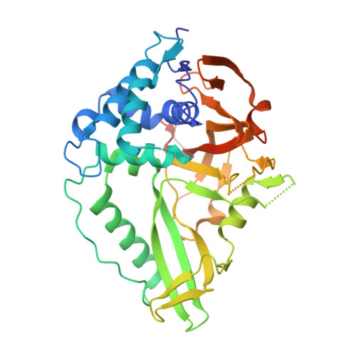
The structure of the deubiquitinase USP15 reveals a misaligned catalytic triad and an open ubiquitin-binding channel.

Ward, S.J.Gratton, H.E.Indrayudha, P.Michavila, C.Mukhopadhyay, R.Maurer, S.K.Caulton, S.G.Emsley, J.Dreveny, I.

(2018) J Biol Chem 293: 17362-17374

  • DOI: https://doi.org/10.1074/jbc.RA118.003857
  • Primary Citation of Related Structures:  
    6GH9, 6GHA

  • PubMed Abstract: 

    Ubiquitin-specific protease 15 (USP15) regulates important cellular processes, including transforming growth factor β (TGF-β) signaling, mitophagy, mRNA processing, and innate immune responses; however, structural information on USP15's catalytic domain is currently unavailable. Here, we determined crystal structures of the USP15 catalytic core domain, revealing a canonical USP fold, including a finger, palm, and thumb region. Unlike for the structure of paralog USP4, the catalytic triad is in an inactive configuration with the catalytic cysteine ∼10 Å apart from the catalytic histidine. This conformation is atypical, and a similar misaligned catalytic triad has so far been observed only for USP7, although USP15 and USP7 are differently regulated. Moreover, we found that the active-site loops are flexible, resulting in a largely open ubiquitin tail-binding channel. Comparison of the USP15 and USP4 structures points to a possible activation mechanism. Sequence differences between these two USPs mainly map to the S1' region likely to confer specificity, whereas the S1 ubiquitin-binding pocket is highly conserved. Isothermal titration calorimetry monoubiquitin- and linear diubiquitin-binding experiments showed significant differences in their thermodynamic profiles, with USP15 displaying a lower affinity for monoubiquitin than USP4. Moreover, we report that USP15 is weakly inhibited by the antineoplastic agent mitoxantrone in vitro A USP15-mitoxantrone complex structure disclosed that the anthracenedione interacts with the S1' binding site. Our results reveal first insights into USP15's catalytic domain structure, conformational changes, differences between paralogs, and small-molecule interactions and establish a framework for cellular probe and inhibitor development.


  • Organizational Affiliation

    From the Centre for Biomolecular Sciences, School of Pharmacy, University of Nottingham, Nottingham NG7 2RD, United Kingdom.


Macromolecules
Find similar proteins by:  (by identity cutoff)  |  3D Structure
Entity ID: 1
MoleculeChains Sequence LengthOrganismDetailsImage
Ubiquitin carboxyl-terminal hydrolase 15
A, B
365Homo sapiensMutation(s): 0 
Gene Names: USP15KIAA0529
EC: 3.4.19.12
UniProt & NIH Common Fund Data Resources
Find proteins for Q9Y4E8 (Homo sapiens)
Explore Q9Y4E8 
Go to UniProtKB:  Q9Y4E8
PHAROS:  Q9Y4E8
GTEx:  ENSG00000135655 
Entity Groups  
Sequence Clusters30% Identity50% Identity70% Identity90% Identity95% Identity100% Identity
UniProt GroupQ9Y4E8
Sequence Annotations
Expand
  • Reference Sequence
Small Molecules
Ligands 3 Unique
IDChains Name / Formula / InChI Key2D Diagram3D Interactions
MIX
Query on MIX

Download Ideal Coordinates CCD File 
E [auth A]1,4-DIHYDROXY-5,8-BIS({2-[(2-HYDROXYETHYL)AMINO]ETHYL}AMINO)-9,10-ANTHRACENEDIONE
C22 H28 N4 O6
KKZJGLLVHKMTCM-UHFFFAOYSA-N
DMS
Query on DMS

Download Ideal Coordinates CCD File 
D [auth A]DIMETHYL SULFOXIDE
C2 H6 O S
IAZDPXIOMUYVGZ-UHFFFAOYSA-N
ZN
Query on ZN

Download Ideal Coordinates CCD File 
C [auth A]ZINC ION
Zn
PTFCDOFLOPIGGS-UHFFFAOYSA-N
Experimental Data & Validation

Experimental Data

  • Method: X-RAY DIFFRACTION
  • Resolution: 2.09 Å
  • R-Value Free: 0.252 
  • R-Value Work: 0.203 
  • R-Value Observed: 0.205 
  • Space Group: P 1 21 1
Unit Cell:
Length ( Å )Angle ( ˚ )
a = 62.07α = 90
b = 94.39β = 90.08
c = 63.29γ = 90
Software Package:
Software NamePurpose
PHENIXrefinement
Aimlessdata scaling
PDB_EXTRACTdata extraction
XDSdata reduction
PHASERphasing

Structure Validation

View Full Validation Report


Ligand Structure Quality Assessment 


Entry History & Funding Information

Deposition Data


Funding OrganizationLocationGrant Number
Biotechnology and Biological Sciences Research CouncilUnited KingdomBB/H012656/1

Revision History  (Full details and data files)

  • Version 1.0: 2018-09-26
    Type: Initial release
  • Version 1.1: 2018-10-03
    Changes: Data collection, Database references
  • Version 1.2: 2018-11-21
    Changes: Data collection, Database references
  • Version 1.3: 2024-01-17
    Changes: Data collection, Database references, Refinement description, Structure summary