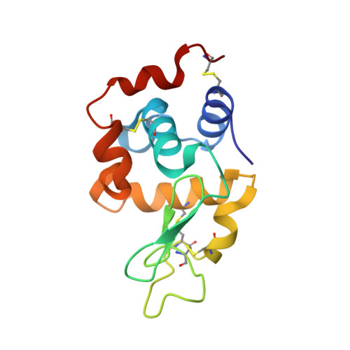
Crystallography on a chip - without the chip: sheet-on-sheet sandwich.
Doak, R.B., Nass Kovacs, G., Gorel, A., Foucar, L., Barends, T.R.M., Grunbein, M.L., Hilpert, M., Kloos, M., Roome, C.M., Shoeman, R.L., Stricker, M., Tono, K., You, D., Ueda, K., Sherrell, D.A., Owen, R.L., Schlichting, I.(2018) Acta Crystallogr D Struct Biol 74: 1000-1007
- PubMed: 30289410
- DOI: https://doi.org/10.1107/S2059798318011634
- Primary Citation of Related Structures:
6GF0, 6HAL - PubMed Abstract:
Crystallography chips are fixed-target supports consisting of a film (for example Kapton) or wafer (for example silicon) that is processed using semiconductor-microfabrication techniques to yield an array of wells or through-holes in which single microcrystals can be lodged for raster-scan probing. Although relatively expensive to fabricate, chips offer an efficient means of high-throughput sample presentation for serial diffraction data collection at synchrotron or X-ray free-electron laser (XFEL) sources. Truly efficient loading of a chip (one microcrystal per well and no wastage during loading) is nonetheless challenging. The wells or holes must match the microcrystal size of interest, requiring that a large stock of chips be maintained. Raster scanning requires special mechanical drives to step the chip rapidly and with micrometre precision from well to well. Here, a `chip-less' adaptation is described that essentially eliminates the challenges of loading and precision scanning, albeit with increased, yet still relatively frugal, sample usage. The device consists simply of two sheets of Mylar with the crystal solution sandwiched between them. This sheet-on-sheet (SOS) sandwich structure has been employed for serial femtosecond crystallography data collection with micrometre-sized crystals at an XFEL. The approach is also well suited to time-resolved pump-probe experiments, in particular for long time delays. The SOS sandwich enables measurements under XFEL beam conditions that would damage conventional chips, as documented here. The SOS sheets hermetically seal the sample, avoiding desiccation of the sample provided that the X-ray beam does not puncture the sheets. This is the case with a synchrotron beam but not with an XFEL beam. In the latter case, desiccation, setting radially outwards from each punched hole, sets lower limits on the speed and line spacing of the raster scan. It is shown that these constraints are easily accommodated.
- Department of Biomolecular Mechanisms, Max Planck Institute for Medical Research, Jahnstrasse 29, 69120 Heidelberg, Germany.
Organizational Affiliation:


