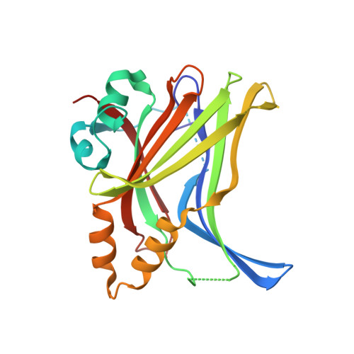
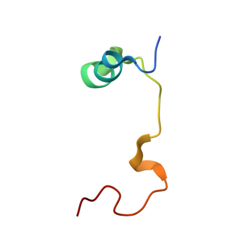
6GEC

TEAD4 (216-434) COMPLEXED WITH YAP PEPTIDE (60-100);S94A AND MYRISTOATE (COVALENTLY BOUND) AT 1.70A (P41212 CRYSTAL FORM); MYRISTOYLATION WAS DONE BY ADDING MYR-COA


Experimental Data Snapshot

  • Method: X-RAY DIFFRACTION
  • Resolution: 1.70 Å
  • R-Value Free: 0.242 
  • R-Value Work: 0.220 
  • R-Value Observed: 0.221 

Starting Model: experimental
View more details

wwPDB Validation   3D Report Full Report


Ligand Structure Quality Assessment 


This is version 1.4 of the entry. See complete history


Literature

Adaptation of the bound intrinsically disordered protein YAP to mutations at the YAP:TEAD interface.

Mesrouze, Y.Bokhovchuk, F.Izaac, A.Meyerhofer, M.Zimmermann, C.Fontana, P.Schmelzle, T.Erdmann, D.Furet, P.Kallen, J.Chene, P.

(2018) Protein Sci 27: 1810-1820

  • DOI: https://doi.org/10.1002/pro.3493
  • Primary Citation of Related Structures:  
    6GE3, 6GE4, 6GE5, 6GE6, 6GEC, 6GEE, 6GEG, 6GEI, 6GEK

  • PubMed Abstract: 

    Many interactions between proteins are mediated by intrinsically disordered regions (IDRs). Intrinsically disordered proteins (IDPs) do not adopt a stable three-dimensional structure in their unbound form, but they become more structured upon binding to their partners. In this communication, we study how a bound IDR adapts to mutations, preventing the formation of hydrogen bonds at the binding interface that needs a precise positioning of the interacting residues to be formed. We use as a model the YAP:TEAD interface, where one YAP (IDP) and two TEAD residues form hydrogen bonds via their side chain. Our study shows that the conformational flexibility of bound YAP and the reorganization of water molecules at the interface help to reduce the energetic constraints created by the loss of H-bonds at the interface. The residual flexibility/dynamic of bound IDRs and water might, therefore, be a key for the adaptation of IDPs to different interface landscapes and to mutations occurring at binding interfaces.


  • Organizational Affiliation

    Disease Area Oncology, Novartis Institutes for Biomedical Research, Basel, Switzerland.


Macromolecules
Find similar proteins by:  (by identity cutoff)  |  3D Structure
Entity ID: 1
MoleculeChains Sequence LengthOrganismDetailsImage
Transcriptional enhancer factor TEF-3219Homo sapiensMutation(s): 0 
Gene Names: TEAD4RTEF1TCF13L1TEF3
UniProt & NIH Common Fund Data Resources
Find proteins for Q15561 (Homo sapiens)
Explore Q15561 
Go to UniProtKB:  Q15561
PHAROS:  Q15561
GTEx:  ENSG00000197905 
Entity Groups  
Sequence Clusters30% Identity50% Identity70% Identity90% Identity95% Identity100% Identity
UniProt GroupQ15561
Sequence Annotations
Expand
  • Reference Sequence
Find similar proteins by:  (by identity cutoff)  |  3D Structure
Entity ID: 2
MoleculeChains Sequence LengthOrganismDetailsImage
Transcriptional coactivator YAP1B [auth L]41Homo sapiensMutation(s): 1 
UniProt & NIH Common Fund Data Resources
Find proteins for P46937 (Homo sapiens)
Explore P46937 
Go to UniProtKB:  P46937
PHAROS:  P46937
GTEx:  ENSG00000137693 
Entity Groups  
Sequence Clusters30% Identity50% Identity70% Identity90% Identity95% Identity100% Identity
UniProt GroupP46937
Sequence Annotations
Expand
  • Reference Sequence
Small Molecules
Ligands 1 Unique
IDChains Name / Formula / InChI Key2D Diagram3D Interactions
MYR
Query on MYR

Download Ideal Coordinates CCD File 
C [auth A]MYRISTIC ACID
C14 H28 O2
TUNFSRHWOTWDNC-UHFFFAOYSA-N
Experimental Data & Validation

Experimental Data

  • Method: X-RAY DIFFRACTION
  • Resolution: 1.70 Å
  • R-Value Free: 0.242 
  • R-Value Work: 0.220 
  • R-Value Observed: 0.221 
  • Space Group: P 41 21 2
Unit Cell:
Length ( Å )Angle ( ˚ )
a = 58.882α = 90
b = 58.882β = 90
c = 158.508γ = 90
Software Package:
Software NamePurpose
XDSdata reduction
XSCALEdata scaling
PHASERphasing
REFMACrefinement
PDB_EXTRACTdata extraction

Structure Validation

View Full Validation Report


Ligand Structure Quality Assessment 


Entry History 

Deposition Data

  • Released Date: 2018-09-19 
  • Deposition Author(s): Kallen, J.

Revision History  (Full details and data files)

  • Version 1.0: 2018-09-19
    Type: Initial release
  • Version 1.1: 2018-10-03
    Changes: Data collection
  • Version 1.2: 2018-10-31
    Changes: Data collection, Database references
  • Version 1.3: 2024-01-17
    Changes: Data collection, Database references, Refinement description
  • Version 1.4: 2024-11-13
    Changes: Structure summary