6GCH

STRUCTURE OF CHYMOTRYPSIN-*TRIFLUOROMETHYL KETONE INHIBITOR COMPLEXES. COMPARISON OF SLOWLY AND RAPIDLY EQUILIBRATING INHIBITORS


Experimental Data Snapshot

  • Method: X-RAY DIFFRACTION
  • Resolution: 2.10 Å
  • R-Value Observed: 0.180 

wwPDB Validation   3D Report Full Report


This is version 1.4 of the entry. See complete history


Literature

Structure of chymotrypsin-trifluoromethyl ketone inhibitor complexes: comparison of slowly and rapidly equilibrating inhibitors.

Brady, K.Wei, A.Z.Ringe, D.Abeles, R.H.

(1990) Biochemistry 29: 7600-7607

  • DOI: https://doi.org/10.1021/bi00485a009
  • Primary Citation of Related Structures:  
    6GCH, 7GCH

  • PubMed Abstract: 

    The peptidyl trifluoromethyl ketones Ac-Phe-CF3 (1) and Ac-Leu-Phe-CF3 (2) are inhibitors of chymotrypsin. They differ in Ki (20 and 2 microM, respectively) as well as in their kinetics of association with chymotrypsin in that 1 is rapidly equilibrating, with an association rate too fast to be observed by steady-state techniques, while 2 is "slow binding", as defined by Morrison and Walsh [Morrison, J. F., & Walsh, C. T. (1988) Adv. Enzymol. Relat. Areas Mol. Biol. 61, 202], with a second-order association rate constant of 750 M-1 s-1 at pH 7.0 [Imperiali, B., & Abeles, R. (1986) Biochemistry 25, 3760]. The crystallographic structures of the complexes of gamma-chymotrypsin with inhibitors 1 and 2 have been determined in order to establish whether structural or conformational differences can be found which account for different kinetic and thermodynamic properties of the two inhibitors. In both complexes, the active-site Ser 195 hydroxyl forms a covalent hemiketal adduct with the trifluoromethyl ketone moiety of the inhibitor. In both complexes, the trifluoromethyl group is partially immobilized, but differences are observed in the degree of interaction of fluorine atoms with the active-site His 57 imidazole ring, with amide nitrogen NH 193, and with other portions of the inhibitor molecule. The enhanced potency of Ac-Leu-Phe-CF3 relative to Ac-Phe-CF3 is accounted for by van der Waals interactions of the leucine side chain of the inhibitor with His 57 and Ile 99 side chains and by a hydrogen bond of the acetyl terminus with amide NH 216 of the enzyme.(ABSTRACT TRUNCATED AT 250 WORDS)


  • Organizational Affiliation

    Department of Toxicology, Harvard School of Public Health, Boston, Massachusetts 02115.


Macromolecules

Find similar proteins by:  Sequence   |   3D Structure  

Entity ID: 1
MoleculeChains Sequence LengthOrganismDetailsImage
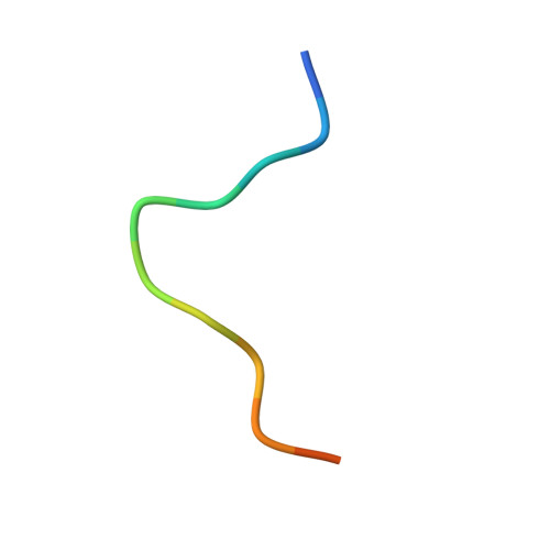
GAMMA-CHYMOTRYPSIN AA [auth E]13Bos taurusMutation(s): 0 
EC: 3.4.21.1
UniProt
Find proteins for P00766 (Bos taurus)
Explore P00766 
Go to UniProtKB:  P00766
Entity Groups  
Sequence Clusters30% Identity50% Identity70% Identity90% Identity95% Identity100% Identity
UniProt GroupP00766
Sequence Annotations
Expand
  • Reference Sequence
Find similar proteins by:  (by identity cutoff)  |  3D Structure
Entity ID: 2
MoleculeChains Sequence LengthOrganismDetailsImage
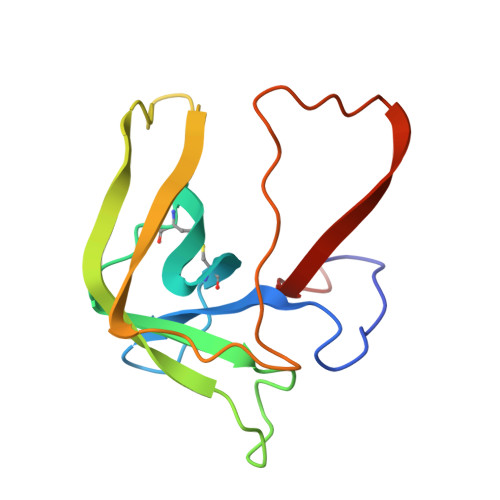
GAMMA-CHYMOTRYPSIN AB [auth F]131Bos taurusMutation(s): 0 
EC: 3.4.21.1
UniProt
Find proteins for P00766 (Bos taurus)
Explore P00766 
Go to UniProtKB:  P00766
Entity Groups  
Sequence Clusters30% Identity50% Identity70% Identity90% Identity95% Identity100% Identity
UniProt GroupP00766
Sequence Annotations
Expand
  • Reference Sequence
Find similar proteins by:  (by identity cutoff)  |  3D Structure
Entity ID: 3
MoleculeChains Sequence LengthOrganismDetailsImage
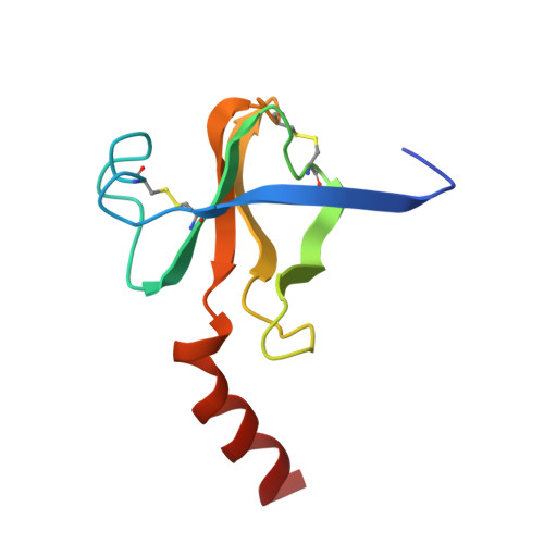
GAMMA-CHYMOTRYPSIN AC [auth G]97Bos taurusMutation(s): 0 
EC: 3.4.21.1
UniProt
Find proteins for P00766 (Bos taurus)
Explore P00766 
Go to UniProtKB:  P00766
Entity Groups  
Sequence Clusters30% Identity50% Identity70% Identity90% Identity95% Identity100% Identity
UniProt GroupP00766
Sequence Annotations
Expand
  • Reference Sequence
Small Molecules
Ligands 1 Unique
IDChains Name / Formula / InChI Key2D Diagram3D Interactions
APF
Query on APF

Download Ideal Coordinates CCD File 
D [auth G]1,1,1-TRIFLUORO-3-ACETAMIDO-4-PHENYL BUTAN-2-ONE(N-ACETYL-L-PHENYLALANYL TRIFLUOROMETHYL KETONE)
C12 H12 F3 N O2
JTIIYPHJIDENCW-JTQLQIEISA-N
Binding Affinity Annotations 
IDSourceBinding Affinity
APF PDBBind:  6GCH Ki: 2.00e+4 (nM) from 1 assay(s)
Experimental Data & Validation

Experimental Data

  • Method: X-RAY DIFFRACTION
  • Resolution: 2.10 Å
  • R-Value Observed: 0.180 
  • Space Group: P 42 21 2
Unit Cell:
Length ( Å )Angle ( ˚ )
a = 69.3α = 90
b = 69.3β = 90
c = 97.2γ = 90
Software Package:
Software NamePurpose
PROLSQrefinement

Structure Validation

View Full Validation Report


Entry History 

Deposition Data

Revision History  (Full details and data files)

  • Version 1.0: 1990-10-15
    Type: Initial release
  • Version 1.1: 2008-03-25
    Changes: Version format compliance
  • Version 1.2: 2011-07-13
    Changes: Version format compliance
  • Version 1.3: 2024-06-05
    Changes: Data collection, Database references, Derived calculations, Other
  • Version 1.4: 2024-11-06
    Changes: Structure summary