6G94 | pdb_00006g94

Structure of E. coli hydrogenase-1 C19G variant in complex with cytochrome b


Experimental Data Snapshot

  • Method: X-RAY DIFFRACTION
  • Resolution: 2.50 Å
  • R-Value Free: 
    0.253 (Depositor), 0.254 (DCC) 
  • R-Value Work: 
    0.222 (Depositor), 0.223 (DCC) 
  • R-Value Observed: 
    0.223 (Depositor) 

Starting Model: experimental
View more details

wwPDB Validation 3D Report Full Report

Validation slider image for 6G94

Ligand Structure Quality Assessment 


This is version 1.5 of the entry. See complete history

Literature

X-ray structural, functional and computational studies of the O2-sensitive E. coli hydrogenase-1 C19G variant reveal an unusual [4Fe-4S] cluster.

Volbeda, A.Mouesca, J.M.Darnault, C.Roessler, M.M.Parkin, A.Armstrong, F.A.Fontecilla-Camps, J.C.

(2018) Chem Commun (Camb) 54: 7175-7178

  • DOI: https://doi.org/10.1039/c8cc02896f
  • Primary Citation Related Structures: 
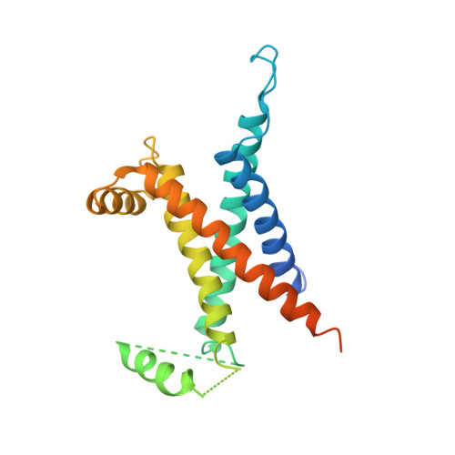
    6G94

  • PubMed Abstract: 

    The crystal structure of the Escherichia coli O2-sensitive C19G [NiFe]-hydrogenase-1 variant shows that the mutation results in a novel FeS cluster, proximal to the Ni-Fe active site. While the proximal cluster of the native O2-tolerant enzyme can transfer two electrons to that site, EPR spectroscopy shows that the modified cluster can transfer only one electron, this shortfall coinciding with O2 sensitivity. Computational studies on electron transfer help to explain how the structural and redox properties of the novel FeS cluster modulate the observed phenotype.


  • Organizational Affiliation
    • Univ. Grenoble Alpes, CEA, CNRS, IBS, F-38000 Grenoble, France. anne.volbeda@ibs.fr.

Macromolecule Content 

  • Total Structure Weight: 467.58 kDa 
  • Atom Count: 30,463 
  • Modeled Residue Count: 3,834 
  • Deposited Residue Count: 4,138 
  • Unique protein chains: 3

Macromolecules

Find similar proteins by:|  3D Structure
Entity ID: 1
MoleculeChains  Sequence LengthOrganismDetailsImage
Hydrogenase-1 small chainA [auth S],
C [auth T],
E [auth Q],
G [auth R]
335Escherichia coli K-12Mutation(s): 0 
Gene Names: hyaAb0972JW0954
EC: 1.12.99.6
Membrane Entity: Yes 
UniProt
Find proteins for P69739 (Escherichia coli (strain K12))
Explore P69739 
Go to UniProtKB:  P69739
Entity Groups
Sequence Clusters30% Identity50% Identity70% Identity90% Identity95% Identity100% Identity
UniProt GroupP69739
Sequence Annotations
Expand
Reference Sequence
Find similar proteins by:|  3D Structure
Entity ID: 2
MoleculeChains  Sequence LengthOrganismDetailsImage
Hydrogenase-1 large chainB [auth L],
D [auth M],
F [auth J],
H [auth K]
582Escherichia coli K-12Mutation(s): 0 
Gene Names: hyaBb0973JW0955
EC: 1.12.99.6
Membrane Entity: Yes 
UniProt
Find proteins for P0ACD8 (Escherichia coli (strain K12))
Explore P0ACD8 
Go to UniProtKB:  P0ACD8
Entity Groups
Sequence Clusters30% Identity50% Identity70% Identity90% Identity95% Identity100% Identity
UniProt GroupP0ACD8
Sequence Annotations
Expand
Reference Sequence
Find similar proteins by:|  3D Structure
Entity ID: 3
MoleculeChains  Sequence LengthOrganismDetailsImage
Probable Ni/Fe-hydrogenase 1 B-type cytochrome subunitI [auth A],
J [auth B]
235Escherichia coli K-12Mutation(s): 0 
Gene Names: hyaCb0974JW0956
Membrane Entity: Yes 
UniProt
Find proteins for P0AAM1 (Escherichia coli (strain K12))
Explore P0AAM1 
Go to UniProtKB:  P0AAM1
Entity Groups
Sequence Clusters30% Identity50% Identity70% Identity90% Identity95% Identity100% Identity
UniProt GroupP0AAM1
Sequence Annotations
Expand
Reference Sequence

Small Molecules

Ligands 8 Unique
IDChains Name / Formula / InChI Key2D Diagram3D Interactions
HEM

Query on HEM


Download:Ideal Coordinates CCD File
MA [auth A],
NA [auth B]
PROTOPORPHYRIN IX CONTAINING FE
C34 H32 Fe N4 O4
KABFMIBPWCXCRK-RGGAHWMASA-L
SF4

Query on SF4


Download:Ideal Coordinates CCD File
FA [auth R],
K [auth S],
R [auth T],
Y [auth Q]
IRON/SULFUR CLUSTER
Fe4 S4
LJBDFODJNLIPKO-UHFFFAOYSA-N
ER2

Query on ER2


Download:Ideal Coordinates CCD File
AA [auth Q],
HA [auth R],
M [auth S],
T
Fe4S4
Fe4 S4
LJBDFODJNLIPKO-UHFFFAOYSA-N
F3S

Query on F3S


Download:Ideal Coordinates CCD File
GA [auth R],
L [auth S],
S [auth T],
Z [auth Q]
FE3-S4 CLUSTER
Fe3 S4
FCXHZBQOKRZXKS-UHFFFAOYSA-N
FCO

Query on FCO


Download:Ideal Coordinates CCD File
BA [auth J],
IA [auth K],
N [auth L],
U [auth M]
CARBONMONOXIDE-(DICYANO) IRON
C3 Fe N2 O
VBQUCMTXYFMTTE-UHFFFAOYSA-N
NI

Query on NI


Download:Ideal Coordinates CCD File
CA [auth J],
JA [auth K],
O [auth L],
V [auth M]
NICKEL (II) ION
Ni
VEQPNABPJHWNSG-UHFFFAOYSA-N
CL

Query on CL


Download:Ideal Coordinates CCD File
EA [auth J],
LA [auth K],
Q [auth L],
X [auth M]
CHLORIDE ION
Cl
VEXZGXHMUGYJMC-UHFFFAOYSA-M
MG

Query on MG


Download:Ideal Coordinates CCD File
DA [auth J],
KA [auth K],
P [auth L],
W [auth M]
MAGNESIUM ION
Mg
JLVVSXFLKOJNIY-UHFFFAOYSA-N

Experimental Data & Validation

Experimental Data

  • Method: X-RAY DIFFRACTION
  • Resolution: 2.50 Å
  • R-Value Free:  0.253 (Depositor), 0.254 (DCC) 
  • R-Value Work:  0.222 (Depositor), 0.223 (DCC) 
  • R-Value Observed: 0.223 (Depositor) 
Space Group: P 21 21 21
Unit Cell:
Length ( Å )Angle ( ˚ )
a = 124.41α = 90
b = 165.03β = 90
c = 206.22γ = 90
Software Package:
Software NamePurpose
XDSdata reduction
Aimlessdata scaling
PHASERphasing
REFMACrefinement
PDB_EXTRACTdata extraction

Structure Validation

View Full Validation Report


Ligand Structure Quality Assessment 


Entry History 

Deposition Data

Revision History  (Full details and data files)

  • Version 1.0: 2018-06-13
    Type: Initial release
  • Version 1.1: 2018-06-20
    Changes: Data collection, Database references
  • Version 1.2: 2018-07-04
    Changes: Data collection, Database references
  • Version 1.3: 2019-08-21
    Changes: Data collection, Database references
  • Version 1.4: 2020-02-05
    Changes: Database references, Source and taxonomy
  • Version 1.5: 2024-01-17
    Changes: Data collection, Database references, Derived calculations, Refinement description