6G5W | pdb_00006g5w

Crystal Structure of KDM4A with compound YP-03-038


Experimental Data Snapshot

  • Method: X-RAY DIFFRACTION
  • Resolution: 1.83 Å
  • R-Value Free: 
    0.218 (Depositor), 0.221 (DCC) 
  • R-Value Work: 
    0.176 (Depositor), 0.191 (DCC) 
  • R-Value Observed: 
    0.177 (Depositor) 

Starting Model: experimental
View more details

wwPDB Validation 3D Report Full Report

Validation slider image for 6G5W

Ligand Structure Quality Assessment 


This is version 1.2 of the entry. See complete history

Literature

Enhanced Properties of a Benzimidazole Benzylpyrazole Lysine Demethylase Inhibitor: Mechanism-of-Action, Binding Site Analysis, and Activity in Cellular Models of Prostate Cancer.

Carter, D.M.Specker, E.Malecki, P.H.Przygodda, J.Dudaniec, K.Weiss, M.S.Heinemann, U.Nazare, M.Gohlke, U.

(2021) J Med Chem 64: 14266-14282

  • DOI: https://doi.org/10.1021/acs.jmedchem.1c00693
  • Primary Citation Related Structures: 
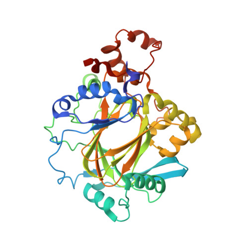
    6G5W, 6G5X

  • PubMed Abstract: 

    Jumonji domain-containing lysine demethylase (KDM) enzymes are encoded by genes of the KDM superfamily. Activities of the KDM4 subfamily promote aggressive phenotypes associated with prostate cancer (PCa). Previously, we discovered a benzimidazole pyrazole molecule that inhibited KDM4 isoforms with properties tractable for development. Here, we demonstrate that a benzyl-substituted variant of this inhibitor exhibits improved potency in biochemical assays, is cell-permeable, and kills PCa cells at low micromolar concentrations. By X-ray crystallography and kinetics-based assays, we demonstrate that the mechanism of inhibition is complex, proceeding via competition with the enzyme for binding of active-site Fe 2+ and by populating a distal site on the enzyme surface. Furthermore, we provide evidence that the inhibitor's cytostatic properties arise from direct intracellular inhibition of KDM4 enzymes. PCa cells treated with the inhibitor exhibit reduced expression of genes regulated by the androgen receptor, an outcome accompanied by epigenetic maintenance of a heterochromatic state.


  • Organizational Affiliation
    • Max Delbrück Center for Molecular Medicine in the Helmholtz Gemeinschaft (MDC), Berlin 13125 Germany.

Macromolecule Content 

  • Total Structure Weight: 84.94 kDa 
  • Atom Count: 6,233 
  • Modeled Residue Count: 692 
  • Deposited Residue Count: 718 
  • Unique protein chains: 1

Macromolecules

Find similar proteins by:|  3D Structure
Entity ID: 1
MoleculeChains  Sequence LengthOrganismDetailsImage
Lysine-specific demethylase 4A
A, B
359Homo sapiensMutation(s): 0 
Gene Names: KDM4AJHDM3AJMJD2JMJD2AKIAA0677
EC: 1.14.11 (PDB Primary Data), 1.14.11.66 (UniProt), 1.14.11.69 (UniProt)
UniProt & NIH Common Fund Data Resources
Find proteins for O75164 (Homo sapiens)
Explore O75164 
Go to UniProtKB:  O75164
PHAROS:  O75164
GTEx:  ENSG00000066135 
Entity Groups
Sequence Clusters30% Identity50% Identity70% Identity90% Identity95% Identity100% Identity
UniProt GroupO75164
Sequence Annotations
Expand
Reference Sequence

Small Molecules

Ligands 6 Unique
IDChains Name / Formula / InChI Key2D Diagram3D Interactions
ENZ

Query on ENZ


Download:Ideal Coordinates CCD File
P [auth B](4~{R})-5-methyl-4-phenyl-2-pyridin-2-yl-pyrazolidin-3-one
C15 H15 N3 O
WTUXNFZMAWODGB-RISCZKNCSA-N
CIT

Query on CIT


Download:Ideal Coordinates CCD File
H [auth A],
I [auth A],
O [auth B]
CITRIC ACID
C6 H8 O7
KRKNYBCHXYNGOX-UHFFFAOYSA-N
ZN

Query on ZN


Download:Ideal Coordinates CCD File
C [auth A],
J [auth B]
ZINC ION
Zn
PTFCDOFLOPIGGS-UHFFFAOYSA-N
EDO

Query on EDO


Download:Ideal Coordinates CCD File
F [auth A],
G [auth A],
N [auth B]
1,2-ETHANEDIOL
C2 H6 O2
LYCAIKOWRPUZTN-UHFFFAOYSA-N
NI

Query on NI


Download:Ideal Coordinates CCD File
D [auth A],
E [auth A],
K [auth B],
L [auth B]
NICKEL (II) ION
Ni
VEQPNABPJHWNSG-UHFFFAOYSA-N
NA

Query on NA


Download:Ideal Coordinates CCD File
M [auth B]SODIUM ION
Na
FKNQFGJONOIPTF-UHFFFAOYSA-N

Experimental Data & Validation

Experimental Data

  • Method: X-RAY DIFFRACTION
  • Resolution: 1.83 Å
  • R-Value Free:  0.218 (Depositor), 0.221 (DCC) 
  • R-Value Work:  0.176 (Depositor), 0.191 (DCC) 
  • R-Value Observed: 0.177 (Depositor) 
Space Group: P 21 21 2
Unit Cell:
Length ( Å )Angle ( ˚ )
a = 99.362α = 90
b = 148.574β = 90
c = 56.39γ = 90
Software Package:
Software NamePurpose
XDSdata reduction
XSCALEdata scaling
PHENIXrefinement
PDB_EXTRACTdata extraction
REFMACphasing

Structure Validation

View Full Validation Report


Ligand Structure Quality Assessment 


Entry History 

Deposition Data

Revision History  (Full details and data files)

  • Version 1.0: 2019-04-10
    Type: Initial release
  • Version 1.1: 2022-12-21
    Changes: Database references, Derived calculations
  • Version 1.2: 2024-01-31
    Changes: Data collection, Refinement description