


Structure of human ADP-forming succinyl-CoA ligase complex SUCLG1-SUCLA2
Bailey, H.J., Shrestha, L., Rembeza, E., Sorrell, F.J., Strain-Damerell, C., Burgess-Brown, N., Arrowsmith, C., Edwards, A., Bountra, C., Yue, W.W.To be published.
Experimental Data Snapshot
Starting Model: experimental
View more details
wwPDB Validation 3D Report Full Report

Entity ID: 1 | |||||
|---|---|---|---|---|---|
| Molecule | Chains | Sequence Length | Organism | Details | Image |
| Succinate--CoA ligase [ADP/GDP-forming] subunit alpha, mitochondrial | 307 | Homo sapiens | Mutation(s): 0 Gene Names: SUCLG1 EC: 6.2.1.4 (PDB Primary Data), 6.2.1.5 (PDB Primary Data), 6.2.1.9 (UniProt), 6.2.1 (UniProt) |  | |
UniProt & NIH Common Fund Data Resources | |||||
PHAROS: P53597 GTEx: ENSG00000163541 | |||||
Entity Groups | |||||
| Sequence Clusters | 30% Identity50% Identity70% Identity90% Identity95% Identity100% Identity | ||||
| UniProt Group | P53597 | ||||
Sequence AnnotationsExpand | |||||
Reference Sequence | |||||
Entity ID: 2 | |||||
|---|---|---|---|---|---|
| Molecule | Chains | Sequence Length | Organism | Details | Image |
| Succinate--CoA ligase [ADP-forming] subunit beta, mitochondrial | 415 | Homo sapiens | Mutation(s): 0 Gene Names: SUCLA2 EC: 6.2.1.5 (PDB Primary Data), 6.2.1.9 (UniProt), 6.2.1 (UniProt) | 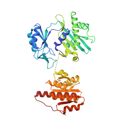 | |
UniProt & NIH Common Fund Data Resources | |||||
PHAROS: Q9P2R7 GTEx: ENSG00000136143 | |||||
Entity Groups | |||||
| Sequence Clusters | 30% Identity50% Identity70% Identity90% Identity95% Identity100% Identity | ||||
| UniProt Group | Q9P2R7 | ||||
Sequence AnnotationsExpand | |||||
Reference Sequence | |||||
| Ligands 1 Unique | |||||
|---|---|---|---|---|---|
| ID | Chains | Name / Formula / InChI Key | 2D Diagram | 3D Interactions | |
| EDO Download:Ideal Coordinates CCD File | C [auth A] D [auth A] E [auth A] F [auth A] G [auth A] | 1,2-ETHANEDIOL C2 H6 O2 LYCAIKOWRPUZTN-UHFFFAOYSA-N |  | ||
| Modified Residues 1 Unique | |||||
|---|---|---|---|---|---|
| ID | Chains | Type | Formula | 2D Diagram | Parent |
| NEP Query on NEP | A | L-PEPTIDE LINKING | C6 H10 N3 O5 P |  | HIS |
| Length ( Å ) | Angle ( ˚ ) |
|---|---|
| a = 130.2 | α = 90 |
| b = 130.2 | β = 90 |
| c = 119.772 | γ = 120 |
| Software Name | Purpose |
|---|---|
| PHENIX | refinement |
| PDB_EXTRACT | data extraction |
| xia2 | data reduction |
| Aimless | data scaling |
| PHASER | phasing |


