6G3W | pdb_00006g3w

Crystal structure of the BIR3 - SERK2 complex from Arabidopsis thaliana.


Experimental Data Snapshot

  • Method: X-RAY DIFFRACTION
  • Resolution: 2.20 Å
  • R-Value Free: 
    0.248 (Depositor), 0.248 (DCC) 
  • R-Value Work: 
    0.217 (Depositor), 0.221 (DCC) 
  • R-Value Observed: 
    0.219 (Depositor) 

Starting Models: experimental
View more details

wwPDB Validation 3D Report Full Report

Validation slider image for 6G3W

Ligand Structure Quality Assessment 


This is version 2.2 of the entry. See complete history

Literature

The SERK3 elongated allele defines a role for BIR ectodomains in brassinosteroid signalling.

Hohmann, U.Nicolet, J.Moretti, A.Hothorn, L.A.Hothorn, M.

(2018) Nat Plants 4: 345-351

  • DOI: https://doi.org/10.1038/s41477-018-0150-9
  • Primary Citation Related Structures: 
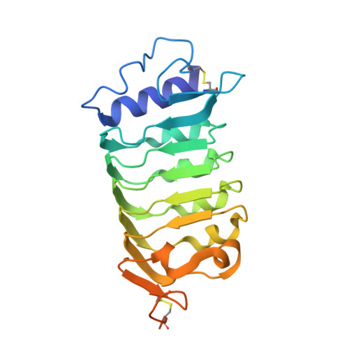
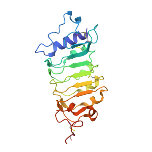
    6FG7, 6FG8, 6G3W

  • PubMed Abstract: 

    The leucine-rich repeat receptor kinase (LRR-RK) BRASSINOSTEROID INSENSITIVE 1 (BRI1) requires a shape-complementary SOMATIC EMBRYOGENESIS RECEPTOR KINASE (SERK) co-receptor for brassinosteroid sensing and receptor activation 1 . Interface mutations that weaken the interaction between receptor and co-receptor in vitro reduce brassinosteroid signalling responses 2 . The SERK3 elongated (elg) allele 3-5 maps to the complex interface and shows enhanced brassinosteroid signalling, but surprisingly no tighter binding to the BRI1 ectodomain in vitro. Here, we report that rather than promoting the interaction with BRI1, the elg mutation disrupts the ability of the co-receptor to interact with the ectodomains of BRI1-ASSOCIATED-KINASE1 INTERACTING KINASE (BIR) receptor pseudokinases, negative regulators of LRR-RK signalling 6 . A conserved lateral surface patch in BIR LRR domains is required for targeting SERK co-receptors and the elg allele maps to the core of the complex interface in a 1.25 Å BIR3-SERK1 structure. Collectively, our structural, quantitative biochemical and genetic analyses suggest that brassinosteroid signalling complex formation is negatively regulated by BIR receptor ectodomains.


  • Organizational Affiliation
    • Structural Plant Biology Laboratory, Department of Botany and Plant Biology, University of Geneva, Geneva, Switzerland.

Macromolecule Content 

  • Total Structure Weight: 92.94 kDa 
  • Atom Count: 5,958 
  • Modeled Residue Count: 746 
  • Deposited Residue Count: 826 
  • Unique protein chains: 2

Macromolecules

Find similar proteins by:|  3D Structure
Entity ID: 1
MoleculeChains  Sequence LengthOrganismDetailsImage
Somatic embryogenesis receptor kinase 2
A, C
217Arabidopsis thalianaMutation(s): 0 
Gene Names: SERK2At1g34210F23M19.11
EC: 2.7.11.1
UniProt
Find proteins for Q9XIC7 (Arabidopsis thaliana)
Explore Q9XIC7 
Go to UniProtKB:  Q9XIC7
Entity Groups
Sequence Clusters30% Identity50% Identity70% Identity90% Identity95% Identity100% Identity
UniProt GroupQ9XIC7
Glycosylation
Glycosylation Sites: 3
Sequence Annotations
Expand
Reference Sequence
Find similar proteins by:|  3D Structure
Entity ID: 2
MoleculeChains  Sequence LengthOrganismDetailsImage
Probable inactive receptor kinase At1g27190
B, D
196Arabidopsis thalianaMutation(s): 0 
Gene Names: At1g27190T7N9.25
UniProt
Find proteins for O04567 (Arabidopsis thaliana)
Explore O04567 
Go to UniProtKB:  O04567
Entity Groups
Sequence Clusters30% Identity50% Identity70% Identity90% Identity95% Identity100% Identity
UniProt GroupO04567
Glycosylation
Glycosylation Sites: 1
Sequence Annotations
Expand
Reference Sequence

Oligosaccharides

Help  
Entity ID: 3
MoleculeChains Length2D Diagram GlycosylationD Interactions
beta-D-mannopyranose-(1-4)-2-acetamido-2-deoxy-beta-D-glucopyranose-(1-4)-2-acetamido-2-deoxy-beta-D-glucopyranose
E, F
3N-Glycosylation
Glycosylation Resources
GlyTouCan: G15407YE
GlyCosmos: G15407YE
GlyGen: G15407YE

Small Molecules

Ligands 3 Unique
IDChains Name / Formula / InChI Key2D Diagram3D Interactions
NAG

Query on NAG


Download:Ideal Coordinates CCD File
G [auth A]
I [auth B]
K [auth C]
L [auth C]
M [auth C]
G [auth A],
I [auth B],
K [auth C],
L [auth C],
M [auth C],
O [auth D]
2-acetamido-2-deoxy-beta-D-glucopyranose
C8 H15 N O6
OVRNDRQMDRJTHS-FMDGEEDCSA-N
PEG

Query on PEG


Download:Ideal Coordinates CCD File
H [auth A]DI(HYDROXYETHYL)ETHER
C4 H10 O3
MTHSVFCYNBDYFN-UHFFFAOYSA-N
EDO

Query on EDO


Download:Ideal Coordinates CCD File
J [auth B],
N [auth C]
1,2-ETHANEDIOL
C2 H6 O2
LYCAIKOWRPUZTN-UHFFFAOYSA-N

Experimental Data & Validation

Experimental Data

  • Method: X-RAY DIFFRACTION
  • Resolution: 2.20 Å
  • R-Value Free:  0.248 (Depositor), 0.248 (DCC) 
  • R-Value Work:  0.217 (Depositor), 0.221 (DCC) 
  • R-Value Observed: 0.219 (Depositor) 
Space Group: P 21 21 21
Unit Cell:
Length ( Å )Angle ( ˚ )
a = 50.184α = 90
b = 52.155β = 90
c = 308.892γ = 90
Software Package:
Software NamePurpose
REFMACrefinement
XDSdata reduction
XDSdata scaling
PHASERphasing

Structure Validation

View Full Validation Report


Ligand Structure Quality Assessment 


Entry History 

& Funding Information

Deposition Data


Funding OrganizationLocationGrant Number
Swiss National Science FoundationSwitzerland31003A_176237

Revision History  (Full details and data files)

  • Version 1.0: 2018-04-04
    Type: Initial release
  • Version 1.1: 2018-05-16
    Changes: Data collection, Database references
  • Version 1.2: 2018-09-05
    Changes: Data collection, Database references
  • Version 2.0: 2020-07-29
    Type: Remediation
    Reason: Carbohydrate remediation
    Changes: Advisory, Atomic model, Data collection, Derived calculations, Structure summary
  • Version 2.1: 2024-01-17
    Changes: Data collection, Database references, Refinement description, Structure summary
  • Version 2.2: 2024-10-16
    Changes: Structure summary