6G20 | pdb_00006g20

Crystal structure of a fluorescence optimized bathy phytochrome PAiRFP2 derived from wild-type Agp2 in its functional Meta-F intermediate state.


Experimental Data Snapshot

  • Method: X-RAY DIFFRACTION
  • Resolution: 2.16 Å
  • R-Value Free: 
    0.232 (Depositor), 0.236 (DCC) 
  • R-Value Work: 
    0.209 (Depositor), 0.213 (DCC) 
  • R-Value Observed: 
    0.210 (Depositor) 

Starting Model: experimental
View more details

wwPDB Validation 3D Report Full Report

Validation slider image for 6G20

Ligand Structure Quality Assessment 


This is version 1.4 of the entry. See complete history

Literature

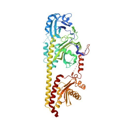
Structural snapshot of a bacterial phytochrome in its functional intermediate state.

Schmidt, A.Sauthof, L.Szczepek, M.Lopez, M.F.Escobar, F.V.Qureshi, B.M.Michael, N.Buhrke, D.Stevens, T.Kwiatkowski, D.von Stetten, D.Mroginski, M.A.Krauss, N.Lamparter, T.Hildebrandt, P.Scheerer, P.

(2018) Nat Commun 9: 4912-4912

  • DOI: https://doi.org/10.1038/s41467-018-07392-7
  • Primary Citation Related Structures: 
    6G1Y, 6G1Z, 6G20

  • PubMed Abstract: 

    Phytochromes are modular photoreceptors of plants, bacteria and fungi that use light as a source of information to regulate fundamental physiological processes. Interconversion between the active and inactive states is accomplished by a photoinduced reaction sequence which couples the sensor with the output module. However, the underlying molecular mechanism is yet not fully understood due to the lack of structural data of functionally relevant intermediate states. Here we report the crystal structure of a Meta-F intermediate state of an Agp2 variant from Agrobacterium fabrum. This intermediate, the identity of which was verified by resonance Raman spectroscopy, was formed by irradiation of the parent Pfr state and displays significant reorientations of almost all amino acids surrounding the chromophore. Structural comparisons allow identifying structural motifs that might serve as conformational switch for initiating the functional secondary structure change that is linked to the (de-)activation of these photoreceptors.


  • Organizational Affiliation
    • Charité - Universitätsmedizin Berlin, Corporate Member of Freie Universität Berlin, Humboldt-Universität zu Berlin, and Berlin Institute of Health, Institute for Medical Physics and Biophysics, Group Protein X-ray Crystallography and Signal Transduction, Charitéplatz 1, Berlin, D-10117, Germany.

Macromolecule Content 

  • Total Structure Weight: 117.71 kDa 
  • Atom Count: 8,259 
  • Modeled Residue Count: 968 
  • Deposited Residue Count: 1,018 
  • Unique protein chains: 1

Macromolecules

Find similar proteins by:|  3D Structure
Entity ID: 1
MoleculeChains  Sequence LengthOrganismDetailsImage
Bacteriophytochrome protein
A, B
509Agrobacterium fabrum str. C58Mutation(s): 24 
Gene Names: SY94_2021
EC: 2.7.13.3
UniProt
Find proteins for A9CI81 (Agrobacterium fabrum (strain C58 / ATCC 33970))
Explore A9CI81 
Go to UniProtKB:  A9CI81
Entity Groups
Sequence Clusters30% Identity50% Identity70% Identity90% Identity95% Identity100% Identity
UniProt GroupA9CI81
Sequence Annotations
Expand
Reference Sequence

Small Molecules

Ligands 7 Unique
IDChains Name / Formula / InChI Key2D Diagram3D Interactions
EL5

Query on EL5


Download:Ideal Coordinates CCD File
C [auth A],
T [auth B]
3-[(2Z)-2-({3-(2-carboxyethyl)-5-[(E)-(4-ethenyl-3-methyl-5-oxo-1,5-dihydro-2H-pyrrol-2-ylidene)methyl]-4-methyl-1H-pyrrol-2-yl}methylidene)-5-{(Z)-[(3E,4S)-3-ethylidene-4-methyl-5-oxopyrrolidin-2-ylidene]methyl}-4-methyl-2H-pyrrol-3-yl]propanoic acid
C33 H36 N4 O6
SNHIGJASYQUMKZ-IDFYGOSVSA-N
ETE

Query on ETE


Download:Ideal Coordinates CCD File
JA [auth B],
S [auth A]
2-{2-[2-2-(METHOXY-ETHOXY)-ETHOXY]-ETHOXY}-ETHANOL
C9 H20 O5
ZNYRFEPBTVGZDN-UHFFFAOYSA-N
PG0

Query on PG0


Download:Ideal Coordinates CCD File
IA [auth B]2-(2-METHOXYETHOXY)ETHANOL
C5 H12 O3
SBASXUCJHJRPEV-UHFFFAOYSA-N
PEG

Query on PEG


Download:Ideal Coordinates CCD File
R [auth A]DI(HYDROXYETHYL)ETHER
C4 H10 O3
MTHSVFCYNBDYFN-UHFFFAOYSA-N
SO4

Query on SO4


Download:Ideal Coordinates CCD File
AA [auth B]
BA [auth B]
CA [auth B]
D [auth A]
DA [auth B]
AA [auth B],
BA [auth B],
CA [auth B],
D [auth A],
DA [auth B],
E [auth A],
F [auth A],
G [auth A],
H [auth A],
I [auth A],
J [auth A],
K [auth A],
L [auth A],
M [auth A],
U [auth B],
V [auth B],
W [auth B],
X [auth B],
Y [auth B],
Z [auth B]
SULFATE ION
O4 S
QAOWNCQODCNURD-UHFFFAOYSA-L
GOL

Query on GOL


Download:Ideal Coordinates CCD File
FA [auth B]
GA [auth B]
HA [auth B]
O [auth A]
P [auth A]
FA [auth B],
GA [auth B],
HA [auth B],
O [auth A],
P [auth A],
Q [auth A]
GLYCEROL
C3 H8 O3
PEDCQBHIVMGVHV-UHFFFAOYSA-N
CL

Query on CL


Download:Ideal Coordinates CCD File
EA [auth B],
N [auth A]
CHLORIDE ION
Cl
VEXZGXHMUGYJMC-UHFFFAOYSA-M

Experimental Data & Validation

Experimental Data

  • Method: X-RAY DIFFRACTION
  • Resolution: 2.16 Å
  • R-Value Free:  0.232 (Depositor), 0.236 (DCC) 
  • R-Value Work:  0.209 (Depositor), 0.213 (DCC) 
  • R-Value Observed: 0.210 (Depositor) 
Space Group: P 63 2 2
Unit Cell:
Length ( Å )Angle ( ˚ )
a = 183.114α = 90
b = 183.114β = 90
c = 179.655γ = 120
Software Package:
Software NamePurpose
REFMACrefinement
XDSdata reduction
SCALAdata scaling
PHASERphasing

Structure Validation

View Full Validation Report


Ligand Structure Quality Assessment 


Entry History 

& Funding Information

Deposition Data


Funding OrganizationLocationGrant Number
GermanySFB1078

Revision History  (Full details and data files)

  • Version 1.0: 2018-11-28
    Type: Initial release
  • Version 1.1: 2018-12-05
    Changes: Data collection, Database references
  • Version 1.2: 2023-05-24
    Changes: Database references, Derived calculations, Structure summary
  • Version 1.3: 2024-02-07
    Changes: Data collection, Refinement description, Structure summary
  • Version 1.4: 2024-10-23
    Changes: Structure summary