6G1G | pdb_00006g1g

GH124 cellulase from Ruminiclostridium thermocellum in complex with Mn and cellotriose


Experimental Data Snapshot

  • Method: X-RAY DIFFRACTION
  • Resolution: 1.04 Å
  • R-Value Free: 
    0.137 (Depositor) 
  • R-Value Work: 
    0.124 (Depositor), 0.124 (DCC) 
  • R-Value Observed: 
    0.125 (Depositor) 

Starting Model: experimental
View more details

wwPDB Validation 3D Report Full Report

Validation slider image for 6G1G

This is version 2.1 of the entry. See complete history

Literature

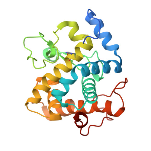
Structural studies of the unusual metal-ion site of the GH124 endoglucanase from Ruminiclostridium thermocellum.

Urresti, S.Cartmell, A.Liu, F.Walton, P.H.Davies, G.J.

(2018) Acta Crystallogr F Struct Biol Commun 74: 496-505

  • DOI: https://doi.org/10.1107/S2053230X18006842
  • Primary Citation Related Structures: 
    6G1G, 6G1I

  • PubMed Abstract: 

    The recent discovery of `lytic' polysaccharide monooxygenases, copper-dependent enzymes for biomass degradation, has provided new impetus for the analysis of unusual metal-ion sites in carbohydrate-active enzymes. In this context, the CAZY family GH124 endoglucanase from Ruminiclostridium thermocellum contains an unusual metal-ion site, which was originally modelled as a Ca 2+ site but features aspartic acid, asparagine and two histidine imidazoles as coordinating residues, which are more consistent with a transition-metal binding environment. It was sought to analyse whether the GH124 metal-ion site might accommodate other metals. It is demonstrated through thermal unfolding experiments that this metal-ion site can accommodate a range of transition metals (Fe 2+ , Cu 2+ , Mn 2+ and Ni 2+ ), whilst the three-dimensional structure and mass spectrometry show that one of the histidines is partially covalently modified and is present as a 2-oxohistidine residue; a feature that is rarely observed but that is believed to be involved in an `off-switch' to transition-metal binding. Atomic resolution (<1.1 Å) complexes define the metal-ion site and also reveal the binding of an unusual fructosylated oligosaccharide, which was presumably present as a contaminant in the cellohexaose used for crystallization. Although it has not been possible to detect a biological role for the unusual metal-ion site, this work highlights the need to study some of the many metal-ion sites in carbohydrate-active enzymes that have long been overlooked or previously mis-assigned.


  • Organizational Affiliation
    • York Structural Biology Laboratory, Department of Chemistry, University of York, York YO10 5DD, England.

Macromolecule Content 

  • Total Structure Weight: 26.41 kDa 
  • Atom Count: 2,168 
  • Modeled Residue Count: 211 
  • Deposited Residue Count: 223 
  • Unique protein chains: 1

Macromolecules

Find similar proteins by:|  3D Structure
Entity ID: 1
MoleculeChains  Sequence LengthOrganismDetailsImage
Glycosyl Hydrolase223Acetivibrio thermocellus ATCC 27405Mutation(s): 0 
Gene Names: Cthe_0435
UniProt
Find proteins for A3DCJ4 (Acetivibrio thermocellus (strain ATCC 27405 / DSM 1237 / JCM 9322 / NBRC 103400 / NCIMB 10682 / NRRL B-4536 / VPI 7372))
Explore A3DCJ4 
Go to UniProtKB:  A3DCJ4
Entity Groups
Sequence Clusters30% Identity50% Identity70% Identity90% Identity95% Identity100% Identity
UniProt GroupA3DCJ4
Sequence Annotations
Expand
Reference Sequence

Oligosaccharides

Help  
Entity ID: 2
MoleculeChains Length2D Diagram GlycosylationD Interactions
beta-D-glucopyranose-(1-4)-beta-D-glucopyranose-(1-4)-beta-D-glucopyranose
B, C
3N/A
Glycosylation Resources
GlyTouCan: G14338VK
GlyCosmos: G14338VK
GlyGen: G14338VK

Small Molecules

Ligands 1 Unique
IDChains Name / Formula / InChI Key2D Diagram3D Interactions
MN
(Subject of Investigation/LOI)

Query on MN


Download:Ideal Coordinates CCD File
D [auth A]MANGANESE (II) ION
Mn
WAEMQWOKJMHJLA-UHFFFAOYSA-N
Modified Residues  1 Unique
IDChains TypeFormula2D DiagramParent
OHI
Query on OHI
A
L-PEPTIDE LINKINGC6 H7 N3 O3HIS

Biologically Interesting Molecules (External Reference) 

1 Unique

Experimental Data & Validation

Experimental Data

  • Method: X-RAY DIFFRACTION
  • Resolution: 1.04 Å
  • R-Value Free:  0.137 (Depositor) 
  • R-Value Work:  0.124 (Depositor), 0.124 (DCC) 
  • R-Value Observed: 0.125 (Depositor) 
Space Group: P 32 2 1
Unit Cell:
Length ( Å )Angle ( ˚ )
a = 73.917α = 90
b = 73.917β = 90
c = 74.936γ = 120
Software Package:
Software NamePurpose
REFMACrefinement
XDSdata reduction
Aimlessdata scaling
MOLREPphasing

Structure Validation

View Full Validation Report


Entry History 

& Funding Information

Deposition Data


Funding OrganizationLocationGrant Number
Biotechnology and Biological Sciences Research CouncilUnited KingdomBB/L021633/1

Revision History  (Full details and data files)

  • Version 1.0: 2018-08-15
    Type: Initial release
  • Version 2.0: 2020-07-29
    Type: Remediation
    Reason: Carbohydrate remediation
    Changes: Atomic model, Data collection, Derived calculations, Structure summary
  • Version 2.1: 2024-01-17
    Changes: Data collection, Database references, Derived calculations, Refinement description, Structure summary