6FPV | pdb_00006fpv

A llama-derived JBP1-targeting nanobody


Experimental Data Snapshot

  • Method: X-RAY DIFFRACTION
  • Resolution: 1.64 Å
  • R-Value Free: 
    0.195 (Depositor), 0.207 (DCC) 
  • R-Value Work: 
    0.163 (Depositor), 0.174 (DCC) 
  • R-Value Observed: 
    0.165 (Depositor) 

Starting Model: experimental
View more details

wwPDB Validation 3D Report Full Report

Validation slider image for 6FPV

This is version 1.3 of the entry. See complete history

Literature

Characterization and structure determination of a llama-derived nanobody targeting the J-base binding protein 1.

van Beusekom, B.Heidebrecht, T.Adamopoulos, A.Fish, A.Pardon, E.Steyaert, J.Joosten, R.P.Perrakis, A.

(2018) Acta Crystallogr F Struct Biol Commun 74: 690-695

  • DOI: https://doi.org/10.1107/S2053230X18010282
  • Primary Citation Related Structures: 
    6FPV

  • PubMed Abstract: 

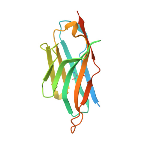
    J-base binding protein 1 (JBP1) contributes to the biosynthesis and maintenance of base J (β-D-glucosylhydroxymethyluracil), a modification of thymidine confined to some protozoa. Camelid (llama) single-domain antibody fragments (nanobodies) targeting JBP1 were produced for use as crystallization chaperones. Surface plasmon resonance screening identified Nb6 as a strong binder, recognizing JBP1 with a 1:1 stoichiometry and high affinity (K d = 30 nM). Crystallization trials of JBP1 in complex with Nb6 yielded crystals that diffracted to 1.47 Å resolution. However, the dimensions of the asymmetric unit and molecular replacement with a nanobody structure clearly showed that the crystals of the expected complex with JBP1 were of the nanobody alone. Nb6 crystallizes in space group P3 1 with two molecules in the asymmetric unit; its crystal structure was refined to a final resolution of 1.64 Å. Ensemble refinement suggests that in the ligand-free state one of the complementarity-determining regions (CDRs) is flexible, while the other two adopt well defined conformations.


  • Organizational Affiliation
    • Department of Biochemistry, Netherlands Cancer Institute, Plesmanlaan 121, 1066 CX Amsterdam, The Netherlands.

Macromolecule Content 

  • Total Structure Weight: 33.55 kDa 
  • Atom Count: 2,119 
  • Modeled Residue Count: 243 
  • Deposited Residue Count: 308 
  • Unique protein chains: 1

Macromolecules

Find similar proteins by:|  3D Structure
Entity ID: 1
MoleculeChains  Sequence LengthOrganismDetailsImage
Nanobody
A, B
154Lama glamaMutation(s): 0 

Experimental Data & Validation

Experimental Data

  • Method: X-RAY DIFFRACTION
  • Resolution: 1.64 Å
  • R-Value Free:  0.195 (Depositor), 0.207 (DCC) 
  • R-Value Work:  0.163 (Depositor), 0.174 (DCC) 
  • R-Value Observed: 0.165 (Depositor) 
Space Group: P 31
Unit Cell:
Length ( Å )Angle ( ˚ )
a = 57.68α = 90
b = 57.68β = 90
c = 64.751γ = 120
Software Package:
Software NamePurpose
XDSdata reduction
Aimlessdata scaling
PHASERphasing
REFMACrefinement
PDB_EXTRACTdata extraction
ARP/wARPmodel building
Cootmodel building

Structure Validation

View Full Validation Report


Entry History 

& Funding Information

Deposition Data


Funding OrganizationLocationGrant Number
NWONetherlandsTOP.13.B2.009
NWONetherlands723.013.003

Revision History  (Full details and data files)

  • Version 1.0: 2018-10-31
    Type: Initial release
  • Version 1.1: 2018-11-14
    Changes: Data collection, Database references
  • Version 1.2: 2024-01-17
    Changes: Data collection, Database references, Refinement description
  • Version 1.3: 2024-11-13
    Changes: Structure summary