6FIJ | pdb_00006fij

Structure of the loading/condensing region (SAT-KS-MAT) of the cercosporin fungal non-reducing polyketide synthase (NR-PKS) CTB1


Experimental Data Snapshot

  • Method: X-RAY DIFFRACTION
  • Resolution: 2.77 Å
  • R-Value Free: 
    0.240 (Depositor), 0.247 (DCC) 
  • R-Value Work: 
    0.205 (Depositor), 0.212 (DCC) 
  • R-Value Observed: 
    0.207 (Depositor) 

Starting Models: experimental
View more details

wwPDB Validation 3D Report Full Report

Validation slider image for 6FIJ

This is version 1.4 of the entry. See complete history

Literature

The structural organization of substrate loading in iterative polyketide synthases.

Herbst, D.A.Huitt-Roehl, C.R.Jakob, R.P.Kravetz, J.M.Storm, P.A.Alley, J.R.Townsend, C.A.Maier, T.

(2018) Nat Chem Biol 14: 474-479

  • DOI: https://doi.org/10.1038/s41589-018-0026-3
  • Primary Citation Related Structures: 
    6FIJ, 6FIK

  • PubMed Abstract: 

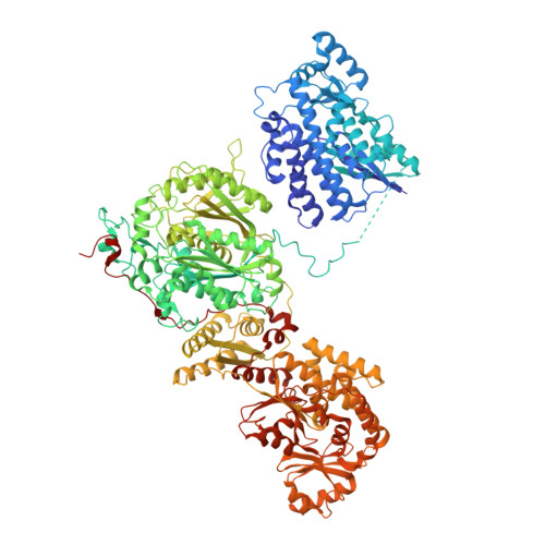
    Polyketide synthases (PKSs) are microbial multienzymes for the biosynthesis of biologically potent secondary metabolites. Polyketide production is initiated by the loading of a starter unit onto an integral acyl carrier protein (ACP) and its subsequent transfer to the ketosynthase (KS). Initial substrate loading is achieved either by multidomain loading modules or by the integration of designated loading domains, such as starter unit acyltransferases (SAT), whose structural integration into PKS remains unresolved. A crystal structure of the loading/condensing region of the nonreducing PKS CTB1 demonstrates the ordered insertion of a pseudodimeric SAT into the condensing region, which is aided by the SAT-KS linker. Cryo-electron microscopy of the post-loading state trapped by mechanism-based crosslinking of ACP to KS reveals asymmetry across the CTB1 loading/-condensing region, in accord with preferential 1:2 binding stoichiometry. These results are critical for re-engineering the loading step in polyketide biosynthesis and support functional relevance of asymmetric conformations of PKSs.


  • Organizational Affiliation
    • Department of Biozentrum, University of Basel, Basel, Switzerland.

Macromolecule Content 

  • Total Structure Weight: 281.71 kDa 
  • Atom Count: 19,867 
  • Modeled Residue Count: 2,550 
  • Deposited Residue Count: 2,608 
  • Unique protein chains: 1

Macromolecules

Find similar proteins by:|  3D Structure
Entity ID: 1
MoleculeChains  Sequence LengthOrganismDetailsImage
Polyketide synthase
A, B
1,304Cercospora nicotianaeMutation(s): 1 
EC: 2.3.1
UniProt
Find proteins for Q6DQW3 (Cercospora nicotianae)
Explore Q6DQW3 
Go to UniProtKB:  Q6DQW3
Entity Groups
Sequence Clusters30% Identity50% Identity70% Identity90% Identity95% Identity100% Identity
UniProt GroupQ6DQW3
Sequence Annotations
Expand
Reference Sequence

Small Molecules

Ligands 4 Unique
IDChains Name / Formula / InChI Key2D Diagram3D Interactions
DTT

Query on DTT


Download:Ideal Coordinates CCD File
R [auth B]2,3-DIHYDROXY-1,4-DITHIOBUTANE
C4 H10 O2 S2
VHJLVAABSRFDPM-IMJSIDKUSA-N
GOL

Query on GOL


Download:Ideal Coordinates CCD File
I [auth A],
J [auth A]
GLYCEROL
C3 H8 O3
PEDCQBHIVMGVHV-UHFFFAOYSA-N
EDO

Query on EDO


Download:Ideal Coordinates CCD File
C [auth A]
D [auth A]
E [auth A]
F [auth A]
G [auth A]
C [auth A],
D [auth A],
E [auth A],
F [auth A],
G [auth A],
H [auth A],
K [auth B],
L [auth B],
M [auth B],
N [auth B],
O [auth B],
P [auth B]
1,2-ETHANEDIOL
C2 H6 O2
LYCAIKOWRPUZTN-UHFFFAOYSA-N
MG

Query on MG


Download:Ideal Coordinates CCD File
Q [auth B]MAGNESIUM ION
Mg
JLVVSXFLKOJNIY-UHFFFAOYSA-N

Experimental Data & Validation

Experimental Data

  • Method: X-RAY DIFFRACTION
  • Resolution: 2.77 Å
  • R-Value Free:  0.240 (Depositor), 0.247 (DCC) 
  • R-Value Work:  0.205 (Depositor), 0.212 (DCC) 
  • R-Value Observed: 0.207 (Depositor) 
Space Group: C 2 2 21
Unit Cell:
Length ( Å )Angle ( ˚ )
a = 108.05α = 90
b = 230.2β = 90
c = 253.8γ = 90
Software Package:
Software NamePurpose
PHENIXrefinement
XDSdata reduction
XSCALEdata scaling
PHASERphasing

Structure Validation

View Full Validation Report


Entry History 

& Funding Information

Deposition Data


Funding OrganizationLocationGrant Number
Swiss National Science FoundationSwitzerland159696
Swiss National Science FoundationSwitzerland145023
Swiss National Science FoundationSwitzerland164074
Swiss National Science FoundationSwitzerland138262
National Institutes of HealthUnited StatesES001670

Revision History  (Full details and data files)

  • Version 1.0: 2018-03-21
    Type: Initial release
  • Version 1.1: 2018-04-04
    Changes: Data collection, Database references, Refinement description
  • Version 1.2: 2018-04-11
    Changes: Data collection, Database references, Derived calculations, Structure summary
  • Version 1.3: 2018-04-25
    Changes: Data collection, Database references, Derived calculations, Structure summary
  • Version 1.4: 2024-01-17
    Changes: Data collection, Database references, Derived calculations, Refinement description