6FHD

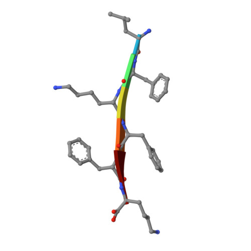
Crystal Structure of the Amyloid-like, out-of-register beta-sheets, polymorph of the LFKFFK segment from the S. aureus PSMalpha3

  • Classification: PROTEIN FIBRIL
  • Organism(s): Staphylococcus aureus
  • Mutation(s): No 

  • Deposited: 2018-01-14 Released: 2018-08-08 
  • Deposition Author(s): Landau, M., Salinas, N.
  • Funding Organization(s): Israel Science Foundation, Deutsch-IsraelischeProjektkooperation (DIP), I-CORE Program of the Planning and Budgeting Committee and The Israel Science Foundation, United States - Israel Binational Science Foundation (BSF)

Experimental Data Snapshot

  • Method: X-RAY DIFFRACTION
  • Resolution: 1.85 Å
  • R-Value Free: 0.187 
  • R-Value Work: 0.171 
  • R-Value Observed: 0.173 

Starting Model: in silico
View more details

wwPDB Validation   3D Report Full Report


This is version 1.3 of the entry. See complete history


Literature

Extreme amyloid polymorphism in Staphylococcus aureus virulent PSM alpha peptides.

Salinas, N.Colletier, J.P.Moshe, A.Landau, M.

(2018) Nat Commun 9: 3512-3512

  • DOI: https://doi.org/10.1038/s41467-018-05490-0
  • Primary Citation of Related Structures:  
    6FG4, 6FGR, 6FHC, 6FHD

  • PubMed Abstract: 

    Members of the Staphylococcus aureus phenol-soluble modulin (PSM) peptide family are secreted as functional amyloids that serve diverse roles in pathogenicity and may be present as full-length peptides or as naturally occurring truncations. We recently showed that the activity of PSMα3, the most toxic member, stems from the formation of cross-α fibrils, which are at variance with the cross-β fibrils linked with eukaryotic amyloid pathologies. Here, we show that PSMα1 and PSMα4, involved in biofilm structuring, form canonical cross-β amyloid fibrils wherein β-sheets tightly mate through steric zipper interfaces, conferring high stability. Contrastingly, a truncated PSMα3 has antibacterial activity, forms reversible fibrils, and reveals two polymorphic and atypical β-rich fibril architectures. These architectures are radically different from both the cross-α fibrils formed by full-length PSMα3, and from the canonical cross-β fibrils. Our results point to structural plasticity being at the basis of the functional diversity exhibited by S. aureus PSMαs.


  • Organizational Affiliation

    Department of Biology, Technion-Israel Institute of Technology, Haifa, 3200003, Israel.


Macromolecules

Find similar proteins by:  Sequence   |   3D Structure  

Entity ID: 1
MoleculeChains Sequence LengthOrganismDetailsImage
Psm alpha-3A [auth B],
B [auth A]
6Staphylococcus aureusMutation(s): 0 
UniProt
Find proteins for H9BRQ7 (Staphylococcus aureus)
Explore H9BRQ7 
Go to UniProtKB:  H9BRQ7
Entity Groups  
UniProt GroupH9BRQ7
Sequence Annotations
Expand
  • Reference Sequence
Experimental Data & Validation

Experimental Data

  • Method: X-RAY DIFFRACTION
  • Resolution: 1.85 Å
  • R-Value Free: 0.187 
  • R-Value Work: 0.171 
  • R-Value Observed: 0.173 
  • Space Group: C 1 2 1
Unit Cell:
Length ( Å )Angle ( ˚ )
a = 41.03α = 90
b = 11.73β = 121.89
c = 24.61γ = 90
Software Package:
Software NamePurpose
XDSdata reduction
XSCALEdata scaling
PHASERphasing
REFMACrefinement
PDB_EXTRACTdata extraction

Structure Validation

View Full Validation Report


Entry History & Funding Information

Deposition Data


Funding OrganizationLocationGrant Number
Israel Science FoundationIsrael560/16
Deutsch-IsraelischeProjektkooperation (DIP)Israel3655/1-1
I-CORE Program of the Planning and Budgeting Committee and The Israel Science FoundationIsrael1775/12
United States - Israel Binational Science Foundation (BSF)Israel2013254

Revision History  (Full details and data files)

  • Version 1.0: 2018-08-08
    Type: Initial release
  • Version 1.1: 2018-09-12
    Changes: Data collection, Database references
  • Version 1.2: 2022-03-30
    Changes: Author supporting evidence, Database references
  • Version 1.3: 2024-05-01
    Changes: Data collection, Refinement description