


The structure of Schistosoma mansoni Phosphodiesterase 4A
Brown, D.G., Schroeder, S., Gil, C., Munday, J., Koning, H., Siderius, M., Leurs, R.To be published.
Experimental Data Snapshot
Starting Model: experimental
View more details
wwPDB Validation 3D Report Full Report

Entity ID: 1 | |||||
|---|---|---|---|---|---|
| Molecule | Chains | Sequence Length | Organism | Details | Image |
| Phosphodiesterase | 370 | Schistosoma mansoni | Mutation(s): 0 Gene Names: Smp_134140 EC: 3.1.4 | 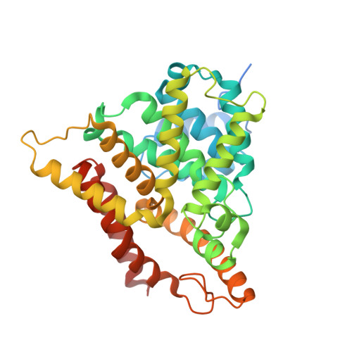 | |
| Ligands 2 Unique | |||||
|---|---|---|---|---|---|
| ID | Chains | Name / Formula / InChI Key | 2D Diagram | 3D Interactions | |
| ZN Download:Ideal Coordinates CCD File | C [auth A], E [auth B] | ZINC ION Zn PTFCDOFLOPIGGS-UHFFFAOYSA-N |  | ||
| MG Download:Ideal Coordinates CCD File | D [auth A], F [auth B] | MAGNESIUM ION Mg JLVVSXFLKOJNIY-UHFFFAOYSA-N |  | ||
| Length ( Å ) | Angle ( ˚ ) |
|---|---|
| a = 81.799 | α = 90 |
| b = 81.799 | β = 90 |
| c = 256.127 | γ = 120 |
| Software Name | Purpose |
|---|---|
| REFMAC | refinement |
| XDS | data reduction |
| Aimless | data scaling |
| MxCuBE | data collection |
| PHASER | phasing |
| Funding Organization | Location | Grant Number |
|---|---|---|
| European Union | United Kingdom | EC-FP7-602666-PDE4NPD |


