Crystal structure of an L chain optimised 14F7 anti-ganglioside Fv suggests a unique tumour-specificity through an unusual H-chain CDR3 architecture.
Bjerregaard-Andersen, K., Johannesen, H., Abdel-Rahman, N., Heggelund, J.E., Hoas, H.M., Abraha, F., Bousquet, P.A., Hoydahl, L.S., Burschowsky, D., Rojas, G., Oscarson, S., Loset, G.A., Krengel, U.(2018) Sci Rep 8: 10836-10836
- PubMed: 30022069 Search on PubMedSearch on PubMed Central
- DOI: https://doi.org/10.1038/s41598-018-28918-5
- Primary Citation Related Structures:
6FFJ - PubMed Abstract:
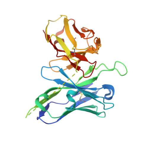
Targeted cancer immunotherapy offers increased efficacy concomitantly with reduced side effects. One antibody with promising clinical potential is 14F7, which specifically recognises the NeuGc GM3 ganglioside. This antigen is found in the plasma membrane of a range of tumours, but is essentially absent from healthy human cells. 14F7 can discriminate NeuGc GM3 from the very similar NeuAc GM3, a common component of cell membranes. The molecular basis for this unique specificity is poorly understood. Here we designed and expressed 14F7-derived single-chain Fvs (scFvs), which retained the specificity of the parent antibody. Detailed expression and purification protocols are described as well as the synthesis of the NeuGc GM3 trisaccharide. The most successful scFv construct, which comprises an alternative variable light chain (V LA ), allowed structure determination to 2.2 Å resolution. The structure gives insights into the conformation of the important CDR H3 loop and the suspected antigen binding site. Furthermore, the presence of V LA instead of the original V L elucidates how this subdomain indirectly stabilises the CDR H3 loop. The current work may serve as a guideline for the efficient production of scFvs for structure determination.
- Department of Chemistry, University of Oslo, NO-0315 Oslo, Norway. kaarebj@kjemi.uio.no.
Organizational Affiliation: