


Aromatic heterocycle galectin-1 interactions for selective single-digit nM affinity ligands
Peterson, A., Collins, P.M., Kahl-Knutsson, B., Zetterberg, F.R., Blanchard, H., Leffler, H., Nilsson, U.J.To be published.
Experimental Data Snapshot
Starting Model: experimental
View more details
Entity ID: 1 | |||||
|---|---|---|---|---|---|
| Molecule | Chains | Sequence Length | Organism | Details | Image |
| Galectin-1 | 134 | Homo sapiens | Mutation(s): 0 Gene Names: LGALS1 | 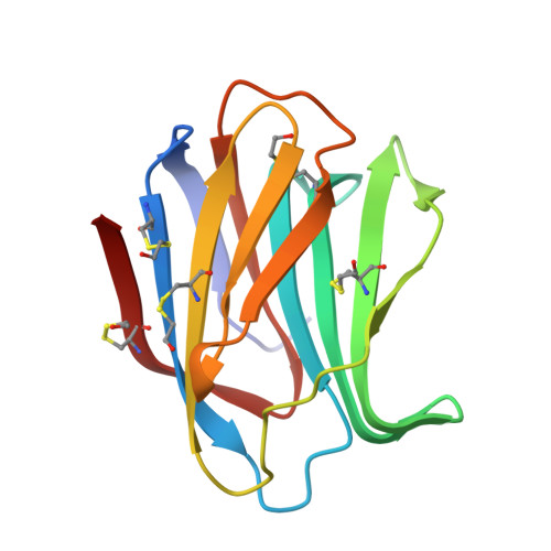 | |
UniProt & NIH Common Fund Data Resources | |||||
PHAROS: P09382 GTEx: ENSG00000100097 | |||||
Entity Groups | |||||
| Sequence Clusters | 30% Identity50% Identity70% Identity90% Identity95% Identity100% Identity | ||||
| UniProt Group | P09382 | ||||
Sequence AnnotationsExpand | |||||
Reference Sequence | |||||
| Ligands 1 Unique | |||||
|---|---|---|---|---|---|
| ID | Chains | Name / Formula / InChI Key | 2D Diagram | 3D Interactions | |
| 5KT (Subject of Investigation/LOI) Download:Ideal Coordinates CCD File | C [auth A], D [auth B] | 3-deoxy-3-[4-(thiophen-3-yl)-1H-1,2,3-triazol-1-yl]-beta-D-galactopyranosyl 3-deoxy-1-thio-3-[4-(thiophen-3-yl)-1H-1,2,3-triazol-1-yl]-beta-D-galactopyranoside C24 H28 N6 O8 S3 OYKAQKLJBYMZSV-VLLPDFJVSA-N |  | ||
| Modified Residues 1 Unique | |||||
|---|---|---|---|---|---|
| ID | Chains | Type | Formula | 2D Diagram | Parent |
| CME Query on CME | A, B | L-PEPTIDE LINKING | C5 H11 N O3 S2 |  | CYS |
| Length ( Å ) | Angle ( ˚ ) |
|---|---|
| a = 44.447 | α = 90 |
| b = 58.689 | β = 90 |
| c = 112.353 | γ = 90 |
| Software Name | Purpose |
|---|---|
| SCALA | data scaling |
| REFMAC | refinement |
| PDB_EXTRACT | data extraction |
| PROTEUM PLUS | data reduction |
| REFMAC | phasing |
| Funding Organization | Location | Grant Number |
|---|---|---|
| Cancer Council Queensland | Australia | ID1080845 |


