6F6L | pdb_00006f6l

R2-like ligand-binding oxidase Y162F mutant with aerobically reconstituted Mn/Fe cofactor


Experimental Data Snapshot

  • Method: X-RAY DIFFRACTION
  • Resolution: 1.90 Å
  • R-Value Free: 
    0.204 (Depositor), 0.209 (DCC) 
  • R-Value Work: 
    0.166 (Depositor), 0.177 (DCC) 
  • R-Value Observed: 
    0.168 (Depositor) 

Starting Model: experimental
View more details

wwPDB Validation 3D Report Full Report

Validation slider image for 6F6L

Ligand Structure Quality Assessment 


This is version 1.3 of the entry. See complete history

Literature

Assembly of a heterodinuclear Mn/Fe cofactor is coupled to tyrosine-valine ether cross-link formation in the R2-like ligand-binding oxidase.

Griese, J.J.Kositzki, R.Haumann, M.Hogbom, M.

(2019) J Biol Inorg Chem 24: 211-221

  • DOI: https://doi.org/10.1007/s00775-019-01639-4
  • Primary Citation Related Structures: 
    6F65, 6F6B, 6F6L, 6F6M

  • PubMed Abstract: 

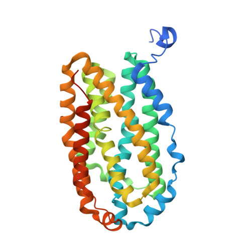
    R2-like ligand-binding oxidases (R2lox) assemble a heterodinuclear Mn/Fe cofactor which performs reductive dioxygen (O 2 ) activation, catalyzes formation of a tyrosine-valine ether cross-link in the protein scaffold, and binds a fatty acid in a putative substrate channel. We have previously shown that the N-terminal metal binding site 1 is unspecific for manganese or iron in the absence of O 2 , but prefers manganese in the presence of O 2 , whereas the C-terminal site 2 is specific for iron. Here, we analyze the effects of amino acid exchanges in the cofactor environment on cofactor assembly and metalation specificity using X-ray crystallography, X-ray absorption spectroscopy, and metal quantification. We find that exchange of either the cross-linking tyrosine or the valine, regardless of whether the mutation still allows cross-link formation or not, results in unspecific manganese or iron binding at site 1 both in the absence or presence of O 2 , while site 2 still prefers iron as in the wild-type. In contrast, a mutation that blocks binding of the fatty acid does not affect the metal specificity of either site under anoxic or aerobic conditions, and cross-link formation is still observed. All variants assemble a dinuclear trivalent metal cofactor in the aerobic resting state, independently of cross-link formation. These findings imply that the cross-link residues are required to achieve the preference for manganese in site 1 in the presence of O 2 . The metalation specificity, therefore, appears to be established during the redox reactions leading to cross-link formation.


  • Organizational Affiliation
    • Department of Biochemistry and Biophysics, Stockholm University, 106 91, Stockholm, Sweden. julia.griese@icm.uu.se.

Macromolecule Content 

  • Total Structure Weight: 74.74 kDa 
  • Atom Count: 4,758 
  • Modeled Residue Count: 556 
  • Deposited Residue Count: 632 
  • Unique protein chains: 1

Macromolecules

Find similar proteins by:|  3D Structure
Entity ID: 1
MoleculeChains  Sequence LengthOrganismDetailsImage
Ribonucleotide reductase small subunit
A, B
316Geobacillus kaustophilus HTA426Mutation(s): 1 
Gene Names: GK2771
EC: 1.17.4.1
UniProt
Find proteins for Q5KW80 (Geobacillus kaustophilus (strain HTA426))
Explore Q5KW80 
Go to UniProtKB:  Q5KW80
Entity Groups
Sequence Clusters30% Identity50% Identity70% Identity90% Identity95% Identity100% Identity
UniProt GroupQ5KW80
Sequence Annotations
Expand
Reference Sequence

Small Molecules

Ligands 4 Unique
IDChains Name / Formula / InChI Key2D Diagram3D Interactions
PLM

Query on PLM


Download:Ideal Coordinates CCD File
F [auth A],
J [auth B]
PALMITIC ACID
C16 H32 O2
IPCSVZSSVZVIGE-UHFFFAOYSA-N
FE

Query on FE


Download:Ideal Coordinates CCD File
E [auth A],
I [auth B]
FE (III) ION
Fe
VTLYFUHAOXGGBS-UHFFFAOYSA-N
MN

Query on MN


Download:Ideal Coordinates CCD File
D [auth A],
H [auth B]
MANGANESE (II) ION
Mn
WAEMQWOKJMHJLA-UHFFFAOYSA-N
MN3

Query on MN3


Download:Ideal Coordinates CCD File
C [auth A],
G [auth B]
MANGANESE (III) ION
Mn
MMIPFLVOWGHZQD-UHFFFAOYSA-N

Experimental Data & Validation

Experimental Data

  • Method: X-RAY DIFFRACTION
  • Resolution: 1.90 Å
  • R-Value Free:  0.204 (Depositor), 0.209 (DCC) 
  • R-Value Work:  0.166 (Depositor), 0.177 (DCC) 
  • R-Value Observed: 0.168 (Depositor) 
Space Group: C 1 2 1
Unit Cell:
Length ( Å )Angle ( ˚ )
a = 162.748α = 90
b = 55.73β = 113.92
c = 69.456γ = 90
Software Package:
Software NamePurpose
PHENIXrefinement
XDSdata reduction
XDSdata scaling
PHASERphasing

Structure Validation

View Full Validation Report


Ligand Structure Quality Assessment 


Entry History 

Deposition Data

Revision History  (Full details and data files)

  • Version 1.0: 2018-12-19
    Type: Initial release
  • Version 1.1: 2019-02-06
    Changes: Data collection, Database references
  • Version 1.2: 2019-03-13
    Changes: Data collection, Database references
  • Version 1.3: 2024-01-17
    Changes: Data collection, Database references, Derived calculations, Refinement description