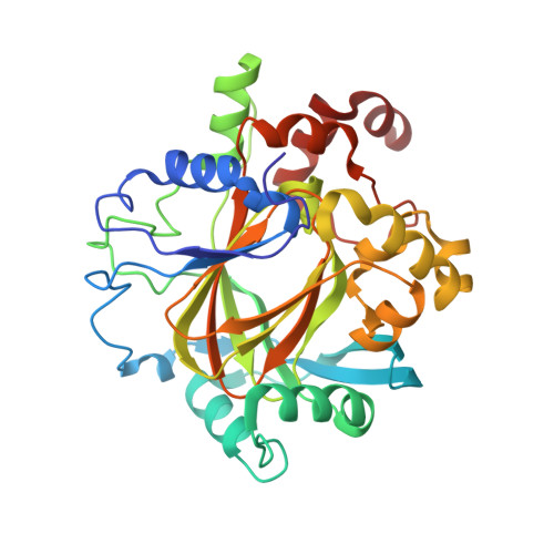
Structure-based mapping of the histone-binding pocket of KDM4D using functionalized tetrazole and pyridine core compounds.
Malecki, P.H., Fassauer, G.M., Ruger, N., Schulig, L., Link, A., Krylova, O., Heinemann, U., Weiss, M.S.(2024) Eur J Med Chem 276: 116642-116642
- PubMed: 38981336 Search on PubMed
- DOI: https://doi.org/10.1016/j.ejmech.2024.116642
- Primary Citation Related Structures:
6F5Q, 6F5R, 6F5S, 6F5T, 6H0W, 6H0X, 6H0Y, 6H0Z, 6H10, 6H11 - PubMed Abstract:
KDM4 histone demethylases became an exciting target for inhibitor development as the evidence linking them directly to tumorigenesis mounts. In this study, we set out to better understand the binding cavity using an X-ray crystallographic approach to provide a detailed landscape of possible interactions within the under-investigated region of KDM4. Our design strategy was based on utilizing known KDM binding motifs, such as nicotinic acid and tetrazolylhydrazides, as core motifs that we decided to enrich with flexible tails to map the distal histone binding site. The resulting X-ray structures of the novel compounds bound to KDM4D, a representative of the KDM4 family, revealed the interaction pattern with distal residues in the histone-binding site. The most prominent protein rearrangement detected upon ligand binding is the loop movement that blocks the accessibility to the histone binding site. Apart from providing new sites that potential inhibitors can target, the novel compounds may prove helpful in exploring the capacity of ligands to bind in sites distal to the cofactor-binding site of other KDMs or 2-oxoglutarate (2OG)-dependent oxygenases. The case study proves that combining a strong small binding motif with flexible tails to probe the binding pocket will facilitate lead discovery in classical drug-discovery campaigns, given the ease of accessing X-ray quality crystals.
- Macromolecular Structure and Interaction, Max-Delbrück-Center for Molecular Medicine, Robert-Rössle-Str. 10, 13125, Berlin, Germany; Macromolecular Crystallography, Helmholtz-Zentrum Berlin für Materialien und Energie, Albert-Einstein-Str. 15, 12489, Berlin, Germany; Department of Structural Biology of Prokaryotic Organisms, Institute of Bioorganic Chemistry, Polish Academy of Sciences, Z. Noskowskiego-Str. 12/14, 61-704, Poznań, Poland. Electronic address: pimalecki@ibch.poznan.pl.
Organizational Affiliation: