6F2F | pdb_00006f2f

Crystal structure of Protease 1 from Pyrococcus Horikoshii co-cystallized in presence of 10 mM Tb-Xo4 and ammonium sulfate.


Experimental Data Snapshot

  • Method: X-RAY DIFFRACTION
  • Resolution: 1.65 Å
  • R-Value Free: 
    0.170 (Depositor), 0.154 (DCC) 
  • R-Value Work: 
    0.161 (Depositor), 0.149 (DCC) 
  • R-Value Observed: 
    0.161 (Depositor) 

Starting Model: experimental
View more details

wwPDB Validation 3D Report Full Report

Validation slider image for 6F2F

Ligand Structure Quality Assessment 


This is version 1.4 of the entry. See complete history

Literature

Unveiling the Binding Modes of the Crystallophore, a Terbium-based Nucleating and Phasing Molecular Agent for Protein Crystallography.

Engilberge, S.Riobe, F.Wagner, T.Di Pietro, S.Breyton, C.Franzetti, B.Shima, S.Girard, E.Dumont, E.Maury, O.

(2018) Chemistry 24: 9739-9746

  • DOI: https://doi.org/10.1002/chem.201802172
  • Primary Citation Related Structures: 
    6F2F, 6F2H, 6F2I, 6F2J, 6F2K, 6F2M, 6FRM, 6FRN, 6FRO, 6FRQ

  • PubMed Abstract: 

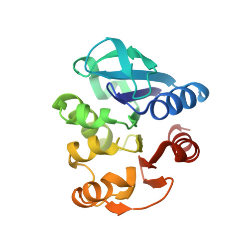
    Crystallophores are lanthanide complexes that act as powerful auxiliary for protein crystallography due to their strong nucleating and phasing effects. To get first insights on the mechanisms behind nucleation induced by Crystallophore, we systematically identified various elaborated networks of supramolecular interactions between Tb-Xo4 and subset of 6 protein structures determined by X-ray diffraction in complex with terbium-Crystallophore (Tb-Xo4). Such interaction mapping analyses demonstrate the versatile binding behavior of the Crystallophore and pave the way to a better understanding of its unique properties.


  • Organizational Affiliation
    • Univ Grenoble Alpes, CEA, CNRS, IBS, 38000, Grenoble, France.

Macromolecule Content 

  • Total Structure Weight: 58.64 kDa 
  • Atom Count: 4,731 
  • Modeled Residue Count: 498 
  • Deposited Residue Count: 498 
  • Unique protein chains: 1

Macromolecules

Find similar proteins by:|  3D Structure
Entity ID: 1
MoleculeChains  Sequence LengthOrganismDetailsImage
Deglycase PH1704
A, B, C
166Pyrococcus horikoshiiMutation(s): 0 
Gene Names: PH1704
EC: 3.5.1.124 (PDB Primary Data), 3.4.22 (PDB Primary Data)
UniProt
Find proteins for O59413 (Pyrococcus horikoshii (strain ATCC 700860 / DSM 12428 / JCM 9974 / NBRC 100139 / OT-3))
Explore O59413 
Go to UniProtKB:  O59413
Entity Groups
Sequence Clusters30% Identity50% Identity70% Identity90% Identity95% Identity100% Identity
UniProt GroupO59413
Sequence Annotations
Expand
Reference Sequence

Small Molecules

Ligands 4 Unique
IDChains Name / Formula / InChI Key2D Diagram3D Interactions
7MT

Query on 7MT


Download:Ideal Coordinates CCD File
D [auth A],
H [auth B],
L [auth C]
Tb-Xo4
C20 H23 N5 O4 Tb
JWLMJALAUZUFRC-UHFFFAOYSA-L
TB

Query on TB


Download:Ideal Coordinates CCD File
G [auth A]TERBIUM(III) ION
Tb
HKCRVXUAKWXBLE-UHFFFAOYSA-N
SO4

Query on SO4


Download:Ideal Coordinates CCD File
E [auth A]
F [auth A]
I [auth B]
J [auth B]
K [auth B]
E [auth A],
F [auth A],
I [auth B],
J [auth B],
K [auth B],
M [auth C],
N [auth C],
O [auth C]
SULFATE ION
O4 S
QAOWNCQODCNURD-UHFFFAOYSA-L
2HA

Query on 2HA


Download:Ideal Coordinates CCD File
P [auth C]Dihydroxyacetone
C3 H6 O3
RXKJFZQQPQGTFL-UHFFFAOYSA-N

Experimental Data & Validation

Experimental Data

  • Method: X-RAY DIFFRACTION
  • Resolution: 1.65 Å
  • R-Value Free:  0.170 (Depositor), 0.154 (DCC) 
  • R-Value Work:  0.161 (Depositor), 0.149 (DCC) 
  • R-Value Observed: 0.161 (Depositor) 
Space Group: P 41 21 2
Unit Cell:
Length ( Å )Angle ( ˚ )
a = 124.588α = 90
b = 124.588β = 90
c = 128.866γ = 90
Software Package:
Software NamePurpose
BUSTERrefinement
XDSdata reduction
SCALAdata scaling
PHASERphasing

Structure Validation

View Full Validation Report


Ligand Structure Quality Assessment 


Entry History 

& Funding Information

Deposition Data


Funding OrganizationLocationGrant Number
French National Research AgencyFranceANR-13-BS07-0007-01

Revision History  (Full details and data files)

  • Version 1.0: 2018-10-03
    Type: Initial release
  • Version 1.1: 2019-05-29
    Changes: Data collection, Derived calculations, Structure summary
  • Version 1.2: 2019-10-16
    Changes: Data collection
  • Version 1.3: 2020-07-08
    Changes: Derived calculations, Structure summary
  • Version 1.4: 2024-01-17
    Changes: Data collection, Database references, Derived calculations, Refinement description