6EZF | pdb_00006ezf

PDE2 in complex with molecule 5


Experimental Data Snapshot

  • Method: X-RAY DIFFRACTION
  • Resolution: 1.50 Å
  • R-Value Free: 
    0.198 (Depositor), 0.204 (DCC) 
  • R-Value Work: 
    0.151 (Depositor) 
  • R-Value Observed: 
    0.152 (Depositor) 

wwPDB Validation 3D Report Full Report

Validation slider image for 6EZF

Ligand Structure Quality Assessment 


This is version 1.2 of the entry. See complete history

Literature

Predicting Binding Free Energies of PDE2 Inhibitors. The Difficulties of Protein Conformation.

Perez-Benito, L.Keranen, H.van Vlijmen, H.Tresadern, G.

(2018) Sci Rep 8: 4883-4883

  • DOI: https://doi.org/10.1038/s41598-018-23039-5
  • Primary Citation Related Structures: 
    6EZF

  • PubMed Abstract: 

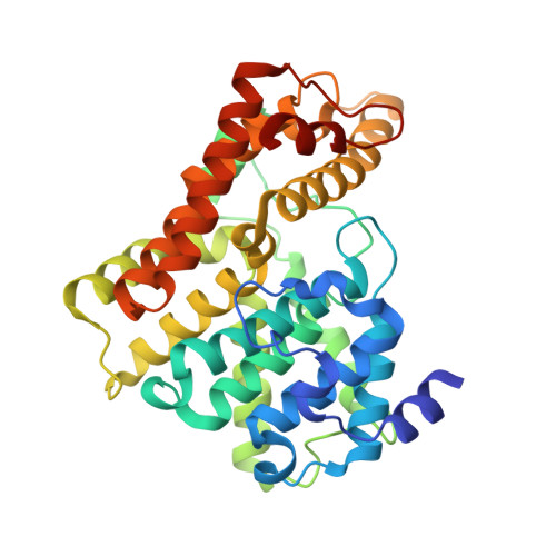
    A congeneric series of 21 phosphodiesterase 2 (PDE2) inhibitors are reported. Crystal structures show how the molecules can occupy a 'top-pocket' of the active site. Molecules with small substituents do not enter the pocket, a critical leucine (Leu770) is closed and water molecules are present. Large substituents enter the pocket, opening the Leu770 conformation and displacing the waters. We also report an X-ray structure revealing a new conformation of the PDE2 active site domain. The relative binding affinities of these compounds were studied with free energy perturbation (FEP) methods and it represents an attractive real-world test case. In general, the calculations could predict the energy of small-to-small, or large-to-large molecule perturbations. However, accurately capturing the transition from small-to-large proved challenging. Only when using alternative protein conformations did results improve. The new X-ray structure, along with a modelled dimer, conferred stability to the catalytic domain during the FEP molecular dynamics (MD) simulations, increasing the convergence and thereby improving the prediction of ΔΔG of binding for some small-to-large transitions. In summary, we found the most significant improvement in results when using different protein structures, and this data set is useful for future free energy validation studies.


  • Organizational Affiliation
    • Computational Chemistry, Janssen Research & Development, Janssen Pharmaceutica N. V., Turnhoutseweg 30, B-2340, Beerse, Belgium.

Macromolecule Content 

  • Total Structure Weight: 42.04 kDa 
  • Atom Count: 3,416 
  • Modeled Residue Count: 342 
  • Deposited Residue Count: 353 
  • Unique protein chains: 1

Macromolecules

Find similar proteins by:|  3D Structure
Entity ID: 1
MoleculeChains  Sequence LengthOrganismDetailsImage
cGMP-dependent 3',5'-cyclic phosphodiesterase353Homo sapiensMutation(s): 0 
EC: 3.1.4.17
UniProt & NIH Common Fund Data Resources
Find proteins for O00408 (Homo sapiens)
Explore O00408 
Go to UniProtKB:  O00408
PHAROS:  O00408
GTEx:  ENSG00000186642 
Entity Groups
Sequence Clusters30% Identity50% Identity70% Identity90% Identity95% Identity100% Identity
UniProt GroupO00408
Sequence Annotations
Expand
Reference Sequence

Small Molecules

Ligands 6 Unique
IDChains Name / Formula / InChI Key2D Diagram3D Interactions
C6Z

Query on C6Z


Download:Ideal Coordinates CCD File
D [auth A]6-[(2,4-dichlorophenyl)methyl]pyridazine-3-thiol
C11 H8 Cl2 N2 S
AOKAGOZDKVLYMU-UHFFFAOYSA-N
SO4

Query on SO4


Download:Ideal Coordinates CCD File
G [auth A]SULFATE ION
O4 S
QAOWNCQODCNURD-UHFFFAOYSA-L
GOL

Query on GOL


Download:Ideal Coordinates CCD File
E [auth A],
F [auth A]
GLYCEROL
C3 H8 O3
PEDCQBHIVMGVHV-UHFFFAOYSA-N
ZN

Query on ZN


Download:Ideal Coordinates CCD File
C [auth A]ZINC ION
Zn
PTFCDOFLOPIGGS-UHFFFAOYSA-N
MG

Query on MG


Download:Ideal Coordinates CCD File
B [auth A]MAGNESIUM ION
Mg
JLVVSXFLKOJNIY-UHFFFAOYSA-N
NA

Query on NA


Download:Ideal Coordinates CCD File
H [auth A]SODIUM ION
Na
FKNQFGJONOIPTF-UHFFFAOYSA-N

Experimental Data & Validation

Experimental Data

  • Method: X-RAY DIFFRACTION
  • Resolution: 1.50 Å
  • R-Value Free:  0.198 (Depositor), 0.204 (DCC) 
  • R-Value Work:  0.151 (Depositor) 
  • R-Value Observed: 0.152 (Depositor) 
Space Group: P 21 21 21
Unit Cell:
Length ( Å )Angle ( ˚ )
a = 54.239α = 90
b = 65.315β = 90
c = 104.391γ = 90
Software Package:
Software NamePurpose
XDSdata reduction
XSCALEdata scaling
REFMACrefinement
PHASERphasing

Structure Validation

View Full Validation Report


Ligand Structure Quality Assessment 


Entry History 

Deposition Data

Revision History  (Full details and data files)

  • Version 1.0: 2018-04-04
    Type: Initial release
  • Version 1.1: 2019-05-08
    Changes: Data collection, Derived calculations
  • Version 1.2: 2024-05-08
    Changes: Advisory, Data collection, Database references