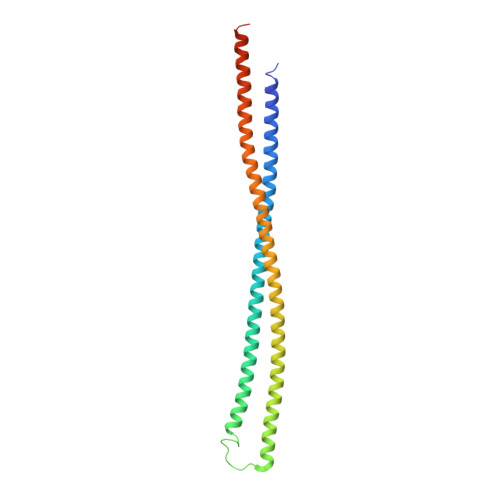
6EWY | pdb_00006ewy

RipA Peptidoglycan hydrolase (Rv1477, Mycobacterium tuberculosis) N-terminal domain


Experimental Data Snapshot

  • Method: X-RAY DIFFRACTION
  • Resolution: 2.20 Å
  • R-Value Free: 
    0.267 (Depositor), 0.275 (DCC) 
  • R-Value Work: 
    0.228 (Depositor), 0.238 (DCC) 
  • R-Value Observed: 
    0.230 (Depositor) 

wwPDB Validation 3D Report Full Report

Validation slider image for 6EWY

This is version 1.3 of the entry. See complete history. 

Literature

The structure of the N-terminal module of the cell wall hydrolase RipA and its role in regulating catalytic activity.

Steiner, E.M., Lyngso, J., Guy, J.E., Bourenkov, G., Lindqvist, Y., Schneider, T.R., Pedersen, J.S., Schneider, G., Schnell, R.

(2018) Proteins 86: 912-923

  • DOI: https://doi.org/10.1002/prot.25523
  • Primary Citation Related Structures: 
    6EWY

  • PubMed Abstract: 

    RipA plays a vital role during cell division of Mycobacterium tuberculosis by degrading the cell wall peptidoglycan at the septum, allowing daughter cell separation. The peptidoglycan degrading activity relies on the NlpC/P60 domain, and as it is potentially harmful when deregulated, spatial and temporal control is necessary in this process. The N-terminal domain of RipA has been proposed to play an inhibitory role blocking the C-terminal NlpC/P60 domain. Accessibility of the active site cysteine residue is however not limited by the presence of the N-terminal domain, but by the lid-module of the inter-domain linker, which is situated in the peptide binding groove of the crystal structures of the catalytic domain. The 2.2 Å resolution structure of the N-terminal domain, determined by Se-SAD phasing, reveals an all-α-fold with 2 long α-helices, and shows similarity to bacterial periplasmic protein domains with scaffold-building role. Size exclusion chromatography and SAXS experiments are consistent with dimer formation of this domain in solution. The SAXS data from the periplasmic two-domain RipA construct suggest a rigid baton-like structure of the N-terminal module, with the catalytic domain connected by a 24 residue long flexible linker. This flexible linker allows for a catalytic zone, which is part of the spatiotemporal control of peptidoglycan degradation.


  • Organizational Affiliation: 
    • Department of Medical Biochemistry and Biophysics, Karolinska Institutet, Stockholm, S-17 177, Sweden.

Macromolecule Content 

  • Total Structure Weight: 23.27 kDa 
  • Atom Count: 1,596 
  • Modeled Residue Count: 201 
  • Deposited Residue Count: 219 
  • Unique protein chains: 1

Macromolecules

Find similar proteins by:|  3D Structure
Entity ID: 1
MoleculeChains  Sequence LengthOrganismDetailsImage
Peptidoglycan endopeptidase RipA219Mycobacterium tuberculosis H37RvMutation(s): 0 
Gene Names: ripA, Rv1477
EC: 3.4
UniProt
Find proteins for O53168 (Mycobacterium tuberculosis (strain ATCC 25618 / H37Rv))
Explore O53168 
Go to UniProtKB:  O53168
Entity Groups
Sequence Clusters30% Identity50% Identity70% Identity90% Identity95% Identity100% Identity
UniProt GroupO53168
Sequence Annotations
Expand
Reference Sequence

Experimental Data & Validation

Experimental Data

  • Method: X-RAY DIFFRACTION
  • Resolution: 2.20 Å
  • R-Value Free:  0.267 (Depositor), 0.275 (DCC) 
  • R-Value Work:  0.228 (Depositor), 0.238 (DCC) 
  • R-Value Observed: 0.230 (Depositor) 
Space Group: C 2 2 2
Unit Cell:
Length ( Å )Angle ( ˚ )
a = 44.916α = 90
b = 93.96β = 90
c = 138.672γ = 90
Software Package:
Software NamePurpose
PHENIXrefinement
XDSdata reduction
autoPROCdata scaling
SHELXDEphasing

Structure Validation

View Full Validation Report


Entry History 

& Funding Information

Deposition Data


Funding OrganizationLocationGrant Number
RAC-VRSweden--

Revision History  (Full details and data files)

  • Version 1.0: 2018-05-02
    Type: Initial release
  • Version 1.1: 2018-05-16
    Changes: Data collection, Database references
  • Version 1.2: 2018-10-24
    Changes: Data collection, Database references
  • Version 1.3: 2024-05-08
    Changes: Data collection, Database references, Refinement description