6EUQ | pdb_00006euq

MdfA(Q131R/L339E)


Experimental Data Snapshot

  • Method: X-RAY DIFFRACTION
  • Resolution: 2.20 Å
  • R-Value Free: 
    0.248 (Depositor), 0.247 (DCC) 
  • R-Value Work: 
    0.231 (Depositor), 0.234 (DCC) 
  • R-Value Observed: 
    0.232 (Depositor) 

Starting Model: experimental
View more details

wwPDB Validation 3D Report Full Report

Validation slider image for 6EUQ

Ligand Structure Quality Assessment 


This is version 1.3 of the entry. See complete history

Literature

A New Critical Conformational Determinant of Multidrug Efflux by an MFS Transporter.

Zomot, E.Yardeni, E.H.Vargiu, A.V.Tam, H.K.Malloci, G.Ramaswamy, V.K.Perach, M.Ruggerone, P.Pos, K.M.Bibi, E.

(2018) J Mol Biology 430: 1368-1385

  • DOI: https://doi.org/10.1016/j.jmb.2018.02.026
  • Primary Citation Related Structures: 
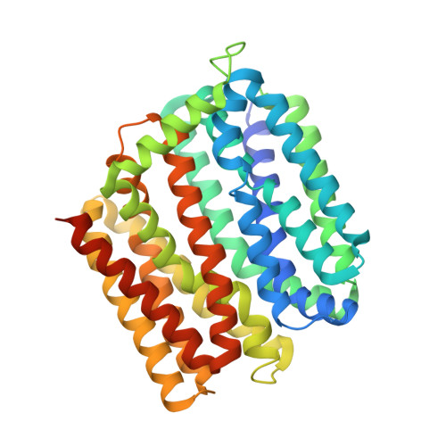
    6EUQ

  • PubMed Abstract: 

    Secondary multidrug (Mdr) transporters utilize ion concentration gradients to actively remove antibiotics and other toxic compounds from cells. The model Mdr transporter MdfA from Escherichia coli exchanges dissimilar drugs for protons. The transporter should open at the cytoplasmic side to enable access of drugs into the Mdr recognition pocket. Here we show that the cytoplasmic rim around the Mdr recognition pocket represents a previously overlooked important regulatory determinant in MdfA. We demonstrate that increasing the positive charge of the electrically asymmetric rim dramatically inhibits MdfA activity and sometimes even leads to influx of planar, positively charged compounds, resulting in drug sensitivity. Our results suggest that unlike the mutants with the electrically modified rim, the membrane-embedded wild-type MdfA exhibits a significant probability of an inward-closed conformation, which is further increased by drug binding. Since MdfA binds drugs from its inward-facing environment, these results are intriguing and raise the possibility that the transporter has a sensitive, drug-induced conformational switch, which favors an inward-closed state.


  • Organizational Affiliation
    • Department of Biomolecular Sciences, Weizmann Institute of Science, Rehovot 76100, Israel.

Macromolecule Content 

  • Total Structure Weight: 47.6 kDa 
  • Atom Count: 3,134 
  • Modeled Residue Count: 386 
  • Deposited Residue Count: 419 
  • Unique protein chains: 1

Macromolecules

Find similar proteins by:|  3D Structure
Entity ID: 1
MoleculeChains  Sequence LengthOrganismDetailsImage
Multidrug transporter MdfA419Escherichia coli K-12Mutation(s): 2 
Gene Names: mdfAcmlAcmrb0842JW0826
Membrane Entity: Yes 
UniProt
Find proteins for P0AEY8 (Escherichia coli (strain K12))
Explore P0AEY8 
Go to UniProtKB:  P0AEY8
Entity Groups
Sequence Clusters30% Identity50% Identity70% Identity90% Identity95% Identity100% Identity
UniProt GroupP0AEY8
Sequence Annotations
Expand
Reference Sequence

Small Molecules

Ligands 8 Unique
IDChains Name / Formula / InChI Key2D Diagram3D Interactions
DXC

Query on DXC


Download:Ideal Coordinates CCD File
C [auth A](3ALPHA,5BETA,12ALPHA)-3,12-DIHYDROXYCHOLAN-24-OIC ACID
C24 H40 O4
KXGVEGMKQFWNSR-LLQZFEROSA-N
BNG

Query on BNG


Download:Ideal Coordinates CCD File
B [auth A]nonyl beta-D-glucopyranoside
C15 H30 O6
QFAPUKLCALRPLH-UXXRCYHCSA-N
PG4

Query on PG4


Download:Ideal Coordinates CCD File
N [auth A]TETRAETHYLENE GLYCOL
C8 H18 O5
UWHCKJMYHZGTIT-UHFFFAOYSA-N
PGE

Query on PGE


Download:Ideal Coordinates CCD File
F [auth A],
G [auth A],
H [auth A]
TRIETHYLENE GLYCOL
C6 H14 O4
ZIBGPFATKBEMQZ-UHFFFAOYSA-N
PR

Query on PR


Download:Ideal Coordinates CCD File
D [auth A],
E [auth A]
PRASEODYMIUM ION
Pr
WCWKKSOQLQEJTE-UHFFFAOYSA-N
OCT

Query on OCT


Download:Ideal Coordinates CCD File
I [auth A],
J [auth A],
K [auth A]
N-OCTANE
C8 H18
TVMXDCGIABBOFY-UHFFFAOYSA-N
HEX

Query on HEX


Download:Ideal Coordinates CCD File
L [auth A]HEXANE
C6 H14
VLKZOEOYAKHREP-UHFFFAOYSA-N
ACT

Query on ACT


Download:Ideal Coordinates CCD File
M [auth A]ACETATE ION
C2 H3 O2
QTBSBXVTEAMEQO-UHFFFAOYSA-M

Experimental Data & Validation

Experimental Data

  • Method: X-RAY DIFFRACTION
  • Resolution: 2.20 Å
  • R-Value Free:  0.248 (Depositor), 0.247 (DCC) 
  • R-Value Work:  0.231 (Depositor), 0.234 (DCC) 
  • R-Value Observed: 0.232 (Depositor) 
Space Group: C 1 2 1
Unit Cell:
Length ( Å )Angle ( ˚ )
a = 95.189α = 90
b = 62.869β = 110.3
c = 100.48γ = 90
Software Package:
Software NamePurpose
REFMACrefinement
XDSdata reduction
Aimlessdata scaling
MOLREPphasing

Structure Validation

View Full Validation Report


Ligand Structure Quality Assessment 


Entry History 

& Funding Information

Deposition Data


Funding OrganizationLocationGrant Number
Germany--
Israel--

Revision History  (Full details and data files)

  • Version 1.0: 2018-08-08
    Type: Initial release
  • Version 1.1: 2018-08-15
    Changes: Data collection, Database references
  • Version 1.2: 2020-07-29
    Type: Remediation
    Reason: Carbohydrate remediation
    Changes: Data collection, Derived calculations, Structure summary
  • Version 1.3: 2024-01-17
    Changes: Data collection, Database references, Refinement description, Structure summary