6ESH

Nucleosome breathing : Class 3


Experimental Data Snapshot

  • Method: ELECTRON MICROSCOPY
  • Resolution: 5.10 Å
  • Aggregation State: PARTICLE 
  • Reconstruction Method: SINGLE PARTICLE 

wwPDB Validation   3D Report Full Report


This is version 1.4 of the entry. See complete history


Literature

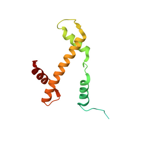
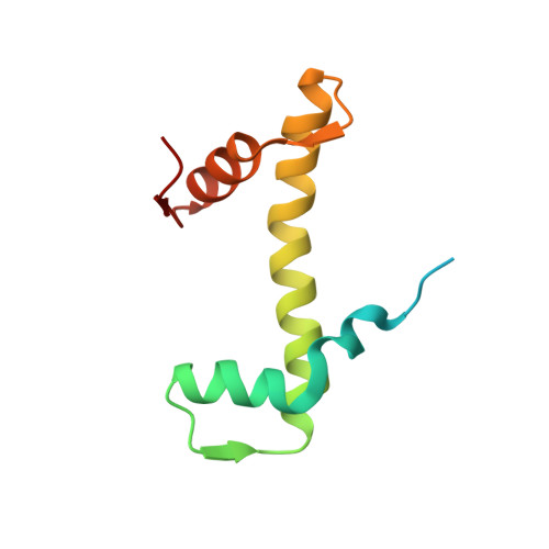
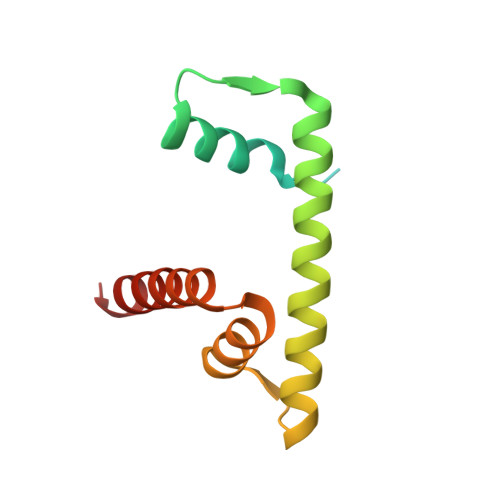
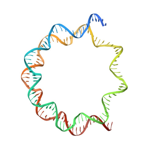
Histone octamer rearranges to adapt to DNA unwrapping.

Bilokapic, S.Strauss, M.Halic, M.

(2018) Nat Struct Mol Biol 25: 101-108

  • DOI: https://doi.org/10.1038/s41594-017-0005-5
  • Primary Citation of Related Structures:  
    6ESF, 6ESG, 6ESH, 6ESI

  • PubMed Abstract: 

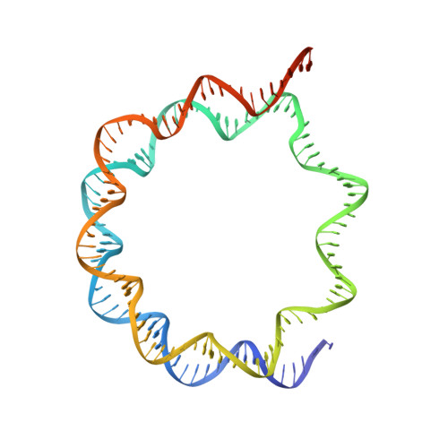
    Nucleosomes, the basic units of chromatin, package and regulate expression of eukaryotic genomes. Although the structure of the intact nucleosome is well characterized, little is known about structures of partially unwrapped, transient intermediates. In this study, we present nine cryo-EM structures of distinct conformations of nucleosome and subnucleosome particles. These structures show that initial DNA breathing induces conformational changes in the histone octamer, particularly in histone H3, that propagate through the nucleosome and prevent symmetrical DNA opening. Rearrangements in the H2A-H2B dimer strengthen interaction with the unwrapping DNA and promote nucleosome stability. In agreement with this, cross-linked H2A-H2B that cannot accommodate unwrapping of the DNA is not stably maintained in the nucleosome. H2A-H2B release and DNA unwrapping occur simultaneously, indicating that DNA is essential in stabilizing the dimer in the nucleosome. Our structures reveal intrinsic nucleosomal plasticity that is required for nucleosome stability and might be exploited by extrinsic protein factors.


  • Organizational Affiliation

    Department of Biochemistry, Gene Center, LMU Munich, Munich, Germany.


Macromolecules

Find similar proteins by:  (by identity cutoff)  |  3D Structure
Entity ID: 1
MoleculeChains Sequence LengthOrganismDetailsImage
Histone H3.2
A, E
135Xenopus laevisMutation(s): 0 
UniProt
Find proteins for P84233 (Xenopus laevis)
Explore P84233 
Go to UniProtKB:  P84233
Entity Groups  
Sequence Clusters30% Identity50% Identity70% Identity90% Identity95% Identity100% Identity
UniProt GroupP84233
Sequence Annotations
Expand
  • Reference Sequence
Find similar proteins by:  (by identity cutoff)  |  3D Structure
Entity ID: 2
MoleculeChains Sequence LengthOrganismDetailsImage
Histone H4
B, F
102Xenopus laevisMutation(s): 0 
UniProt
Find proteins for P62799 (Xenopus laevis)
Explore P62799 
Go to UniProtKB:  P62799
Entity Groups  
Sequence Clusters30% Identity50% Identity70% Identity90% Identity95% Identity100% Identity
UniProt GroupP62799
Sequence Annotations
Expand
  • Reference Sequence
Find similar proteins by:  (by identity cutoff)  |  3D Structure
Entity ID: 3
MoleculeChains Sequence LengthOrganismDetailsImage
Histone H2A
C, G
129Xenopus laevisMutation(s): 0 
Gene Names: hist1h2ajLOC494591
UniProt
Find proteins for Q6AZJ8 (Xenopus laevis)
Explore Q6AZJ8 
Go to UniProtKB:  Q6AZJ8
Entity Groups  
Sequence Clusters30% Identity50% Identity70% Identity90% Identity95% Identity100% Identity
UniProt GroupQ6AZJ8
Sequence Annotations
Expand
  • Reference Sequence
Find similar proteins by:  (by identity cutoff)  |  3D Structure
Entity ID: 4
MoleculeChains Sequence LengthOrganismDetailsImage
Histone H2B 1.1
D, H
122Xenopus laevisMutation(s): 0 
UniProt
Find proteins for P02281 (Xenopus laevis)
Explore P02281 
Go to UniProtKB:  P02281
Entity Groups  
Sequence Clusters30% Identity50% Identity70% Identity90% Identity95% Identity100% Identity
UniProt GroupP02281
Sequence Annotations
Expand
  • Reference Sequence
Find similar nucleic acids by:  (by identity cutoff)  |  3D Structure
Entity ID: 5
MoleculeChains LengthOrganismImage
DNA (137-MER)147synthetic construct
Sequence Annotations
Expand
  • Reference Sequence
Find similar nucleic acids by:  (by identity cutoff)  |  3D Structure
Entity ID: 6
MoleculeChains LengthOrganismImage
DNA (137-MER)147synthetic construct
Sequence Annotations
Expand
  • Reference Sequence
Experimental Data & Validation

Experimental Data

  • Method: ELECTRON MICROSCOPY
  • Resolution: 5.10 Å
  • Aggregation State: PARTICLE 
  • Reconstruction Method: SINGLE PARTICLE 
EM Software:
TaskSoftware PackageVersion
RECONSTRUCTIONRELION2.0

Structure Validation

View Full Validation Report


Entry History & Funding Information

Deposition Data


Funding OrganizationLocationGrant Number
European Research CouncilERC-smallRNAhet-309584

Revision History  (Full details and data files)

  • Version 1.0: 2017-12-20
    Type: Initial release
  • Version 1.1: 2017-12-27
    Changes: Database references
  • Version 1.2: 2018-01-24
    Changes: Database references
  • Version 1.3: 2019-10-02
    Changes: Data collection, Other
  • Version 1.4: 2019-10-23
    Changes: Data collection, Other