6EQL | pdb_00006eql

Crystal Structure of Human Glycogenin-1 (GYG1) Tyr195pIPhe mutant complexed with manganese and UDP


Experimental Data Snapshot

  • Method: X-RAY DIFFRACTION
  • Resolution: 2.38 Å
  • R-Value Free: 
    0.281 (Depositor), 0.280 (DCC) 
  • R-Value Work: 
    0.225 (Depositor), 0.228 (DCC) 
  • R-Value Observed: 
    0.228 (Depositor) 

wwPDB Validation 3D Report Full Report

Validation slider image for 6EQL

Ligand Structure Quality Assessment 


This is version 1.4 of the entry. See complete history

Literature

Palladium-mediated enzyme activation suggests multiphase initiation of glycogenesis.

Bilyard, M.K.Bailey, H.J.Raich, L.Gafitescu, M.A.Machida, T.Iglesias-Fernandez, J.Lee, S.S.Spicer, C.D.Rovira, C.Yue, W.W.Davis, B.G.

(2018) Nature 563: 235-240

  • DOI: https://doi.org/10.1038/s41586-018-0644-7
  • Primary Citation Related Structures: 
    6EQJ, 6EQL

  • PubMed Abstract: 

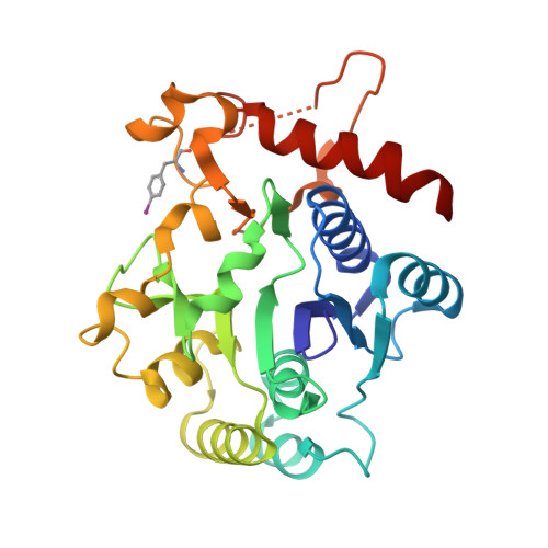
    Biosynthesis of glycogen, the essential glucose (and hence energy) storage molecule in humans, animals and fungi 1 , is initiated by the glycosyltransferase enzyme, glycogenin (GYG). Deficiencies in glycogen formation cause neurodegenerative and metabolic disease 2-4 , and mouse knockout 5 and inherited human mutations 6 of GYG impair glycogen synthesis. GYG acts as a 'seed core' for the formation of the glycogen particle by catalysing its own stepwise autoglucosylation to form a covalently bound gluco-oligosaccharide chain at initiation site Tyr 195. Precise mechanistic studies have so far been prevented by an inability to access homogeneous glycoforms of this protein, which unusually acts as both catalyst and substrate. Here we show that unprecedented direct access to different, homogeneously glucosylated states of GYG can be accomplished through a palladium-mediated enzyme activation 'shunt' process using on-protein C-C bond formation. Careful mimicry of GYG intermediates recapitulates catalytic activity at distinct stages, which in turn allows discovery of triphasic kinetics and substrate plasticity in GYG's use of sugar substrates. This reveals a tolerant but 'proof-read' mechanism that underlies the precision of this metabolic process. The present demonstration of direct, chemically controlled access to intermediate states of active enzymes suggests that such ligation-dependent activation could be a powerful tool in the study of mechanism.


  • Organizational Affiliation
    • Department of Chemistry, University of Oxford, Oxford, UK.

Macromolecule Content 

  • Total Structure Weight: 60.84 kDa 
  • Atom Count: 3,843 
  • Modeled Residue Count: 479 
  • Deposited Residue Count: 526 
  • Unique protein chains: 1

Macromolecules

Find similar proteins by:|  3D Structure
Entity ID: 1
MoleculeChains  Sequence LengthOrganismDetailsImage
Glycogenin-1
A, B
263Homo sapiensMutation(s): 1 
Gene Names: GYG1GYG
EC: 2.4.1.186
UniProt & NIH Common Fund Data Resources
Find proteins for P46976 (Homo sapiens)
Explore P46976 
Go to UniProtKB:  P46976
PHAROS:  P46976
GTEx:  ENSG00000163754 
Entity Groups
Sequence Clusters30% Identity50% Identity70% Identity90% Identity95% Identity100% Identity
UniProt GroupP46976
Sequence Annotations
Expand
Reference Sequence

Small Molecules

Ligands 4 Unique
IDChains Name / Formula / InChI Key2D Diagram3D Interactions
2PE

Query on 2PE


Download:Ideal Coordinates CCD File
G [auth B]NONAETHYLENE GLYCOL
C18 H38 O10
YZUUTMGDONTGTN-UHFFFAOYSA-N
UDP

Query on UDP


Download:Ideal Coordinates CCD File
C [auth A],
E [auth B]
URIDINE-5'-DIPHOSPHATE
C9 H14 N2 O12 P2
XCCTYIAWTASOJW-XVFCMESISA-N
EDO

Query on EDO


Download:Ideal Coordinates CCD File
H [auth B]1,2-ETHANEDIOL
C2 H6 O2
LYCAIKOWRPUZTN-UHFFFAOYSA-N
MN

Query on MN


Download:Ideal Coordinates CCD File
D [auth A],
F [auth B]
MANGANESE (II) ION
Mn
WAEMQWOKJMHJLA-UHFFFAOYSA-N
Modified Residues  1 Unique
IDChains TypeFormula2D DiagramParent
PHI
Query on PHI
A, B
L-PEPTIDE LINKINGC9 H10 I N O2PHE

Experimental Data & Validation

Experimental Data

  • Method: X-RAY DIFFRACTION
  • Resolution: 2.38 Å
  • R-Value Free:  0.281 (Depositor), 0.280 (DCC) 
  • R-Value Work:  0.225 (Depositor), 0.228 (DCC) 
  • R-Value Observed: 0.228 (Depositor) 
Space Group: P 21 21 21
Unit Cell:
Length ( Å )Angle ( ˚ )
a = 66.17α = 90
b = 81.05β = 90
c = 111.2γ = 90
Software Package:
Software NamePurpose
Aimlessdata scaling
PHENIXrefinement
PDB_EXTRACTdata extraction
xia2data reduction
PHASERphasing

Structure Validation

View Full Validation Report


Ligand Structure Quality Assessment 


Entry History 

Deposition Data

Revision History  (Full details and data files)

  • Version 1.0: 2017-12-20
    Type: Initial release
  • Version 1.1: 2018-10-24
    Changes: Data collection, Database references
  • Version 1.2: 2018-11-07
    Changes: Data collection, Database references
  • Version 1.3: 2018-11-14
    Changes: Data collection, Database references
  • Version 1.4: 2025-04-09
    Changes: Data collection, Database references, Derived calculations, Structure summary