6EOL | pdb_00006eol

Human galectin-3c in complex with a galactose derivative


Experimental Data Snapshot

  • Method: X-RAY DIFFRACTION
  • Resolution: 1.50 Å
  • R-Value Free: 
    0.218 (Depositor), 0.215 (DCC) 
  • R-Value Work: 
    0.170 (Depositor), 0.173 (DCC) 
  • R-Value Observed: 
    0.172 (Depositor) 

wwPDB Validation 3D Report Full Report

Validation slider image for 6EOL

Ligand Structure Quality Assessment 


This is version 1.2 of the entry. See complete history

Literature

Monosaccharide Derivatives with Low-Nanomolar Lectin Affinity and High Selectivity Based on Combined Fluorine-Amide, Phenyl-Arginine, Sulfur-pi , and Halogen Bond Interactions.

Zetterberg, F.R.Peterson, K.Johnsson, R.E.Brimert, T.Hakansson, M.Logan, D.T.Leffler, H.Nilsson, U.J.

(2018) ChemMedChem 13: 133-137

  • DOI: https://doi.org/10.1002/cmdc.201700744
  • Primary Citation Related Structures: 
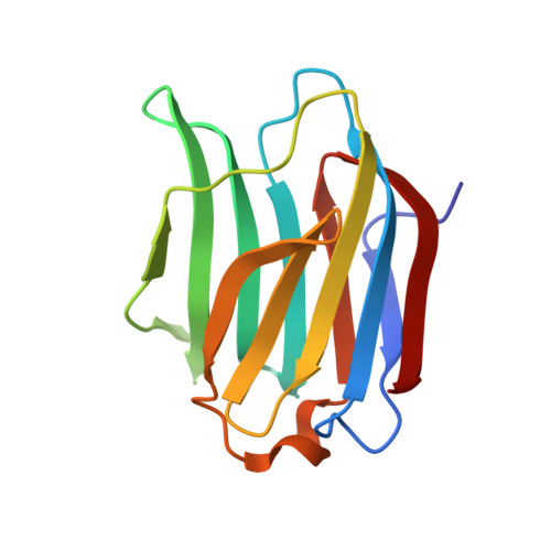
    6EOG, 6EOL

  • PubMed Abstract: 

    The design of small and high-affinity lectin inhibitors remains a major challenge because the natural ligand binding sites of lectin are often shallow and have polar character. Herein we report that derivatizing galactose with un-natural structural elements that form multiple non-natural lectin-ligand interactions (orthogonal multipolar fluorine-amide, phenyl-arginine, sulfur-π, and halogen bond) can provide inhibitors with extraordinary affinity (low nanomolar) for the model lectin, galectin-3, which is more than five orders of magnitude higher than the parent galactose; moreover, is selective over other galectins.


  • Organizational Affiliation
    • Galecto Biotech AB, Sahlgrenska Science Park, Medicinaregatan 8A, 413 46, Gothenburg, Sweden.

Macromolecule Content 

  • Total Structure Weight: 16.37 kDa 
  • Atom Count: 1,538 
  • Modeled Residue Count: 138 
  • Deposited Residue Count: 138 
  • Unique protein chains: 1

Macromolecules

Find similar proteins by:|  3D Structure
Entity ID: 1
MoleculeChains  Sequence LengthOrganismDetailsImage
Galectin-3138Homo sapiensMutation(s): 0 
Gene Names: LGALS3MAC2
UniProt & NIH Common Fund Data Resources
Find proteins for P17931 (Homo sapiens)
Explore P17931 
Go to UniProtKB:  P17931
PHAROS:  P17931
GTEx:  ENSG00000131981 
Entity Groups
Sequence Clusters30% Identity50% Identity70% Identity90% Identity95% Identity100% Identity
UniProt GroupP17931
Sequence Annotations
Expand
Reference Sequence

Small Molecules

Ligands 2 Unique
IDChains Name / Formula / InChI Key2D Diagram3D Interactions
BKH

Query on BKH


Download:Ideal Coordinates CCD File
D [auth A](2~{R},3~{R},4~{S},5~{R},6~{R})-2-(3,4-dichlorophenyl)sulfanyl-6-(hydroxymethyl)-4-[4-[3,4,5-tris(fluoranyl)phenyl]-1,2,3-triazol-1-yl]oxane-3,5-diol
C20 H16 Cl2 F3 N3 O4 S
CSGJIUAIYDKFPC-DABHTEOTSA-N
SCN

Query on SCN


Download:Ideal Coordinates CCD File
B [auth A],
C [auth A]
THIOCYANATE ION
C N S
ZMZDMBWJUHKJPS-UHFFFAOYSA-M

Experimental Data & Validation

Experimental Data

  • Method: X-RAY DIFFRACTION
  • Resolution: 1.50 Å
  • R-Value Free:  0.218 (Depositor), 0.215 (DCC) 
  • R-Value Work:  0.170 (Depositor), 0.173 (DCC) 
  • R-Value Observed: 0.172 (Depositor) 
Space Group: P 21 21 21
Unit Cell:
Length ( Å )Angle ( ˚ )
a = 36.152α = 90
b = 58.26β = 90
c = 62.797γ = 90
Software Package:
Software NamePurpose
BUSTERrefinement
XDSdata reduction
XSCALEdata scaling
REFMACphasing

Structure Validation

View Full Validation Report


Ligand Structure Quality Assessment 


Entry History 

& Funding Information

Deposition Data


Funding OrganizationLocationGrant Number
Swedish Research CouncilSweden621-2012-2978
Knut and Alice Wallenberg FoundationSweden2013.0022

Revision History  (Full details and data files)

  • Version 1.0: 2018-08-22
    Type: Initial release
  • Version 1.1: 2019-10-16
    Changes: Data collection
  • Version 1.2: 2024-05-08
    Changes: Data collection, Database references