6EO9

Crystal structure of thrombin in complex with a novel glucose-conjugated potent inhibitor


Experimental Data Snapshot

  • Method: X-RAY DIFFRACTION
  • Resolution: 1.84 Å
  • R-Value Free: 
    0.248 (Depositor), 0.250 (DCC) 
  • R-Value Work: 
    0.198 (Depositor), 0.200 (DCC) 
  • R-Value Observed: 
    0.201 (Depositor) 

wwPDB Validation   3D Report Full Report


Ligand Structure Quality Assessment 

Created with Raphaël 2.3.0Worse 01 BetterLigand structure goodness of fit to experimental dataBest fitted 2OJClick on this verticalbar to view details

This is version 1.3 of the entry. See complete history


Literature

How a beta-D-glucoside side chain enhances binding affinity to thrombin of inhibitors bearing 2-chlorothiophene as P1 moiety: crystallography, fragment deconstruction study, and evaluation of antithrombotic properties.

Belviso, B.D.Caliandro, R.de Candia, M.Zaetta, G.Lopopolo, G.Incampo, F.Colucci, M.Altomare, C.D.

(2014) J Med Chem 57: 8563-8575

  • DOI: https://doi.org/10.1021/jm5010754
  • Primary Citation of Related Structures:  
    6EO8, 6EO9

  • PubMed Abstract: 

    The β-d-glucose-containing compound 3, bearing 2-chlorothiophene and 1-isopropylpiperidine moieties as binders of the S1 and S4 pockets, respectively, proved to be potent competitive inhibitor of factor Xa (fXa, Ki = 0.090 nM) and thrombin (fIIa, Ki = 100 nM). The potency of 3 increases, over the parent compound 1, against fIIa (110-fold), much more than against fXa (7-fold). Experimental deconstruction of 3 into smaller fragments revealed a binding cooperativity of the P3/P4 and propylene-linked β-d-glucose fragments, stronger in fIIa (15.5 kJ·mol(-1)) than in fXa (2.8 kJ·mol(-1)). The crystal structure of human fIIa in complex with 3 revealed a binding mode including a strong H-bond network between the glucose O1', O3', and O5' and two critical residues, namely R221a and K224, belonging to the Na(+)-binding site which may allosterically perturb the specificity sites. The potential of 3 as antithrombotic agent was supported by its ability to inhibit thrombin generation and to stimulate fibrinolysis at submicromolar concentration.


  • Organizational Affiliation

    Institute of Crystallography, Consiglio Nazionale delle Ricerche , Via Amendola 122/o, 70126 Bari, Italy.


Macromolecules
Find similar proteins by:  (by identity cutoff)  |  3D Structure
Entity ID: 1
MoleculeChains Sequence LengthOrganismDetailsImage
ProthrombinA [auth H]259Homo sapiensMutation(s): 0 
Gene Names: F2
EC: 3.4.21.5
UniProt & NIH Common Fund Data Resources
Find proteins for P00734 (Homo sapiens)
Explore P00734 
Go to UniProtKB:  P00734
PHAROS:  P00734
GTEx:  ENSG00000180210 
Entity Groups  
Sequence Clusters30% Identity50% Identity70% Identity90% Identity95% Identity100% Identity
UniProt GroupP00734
Sequence Annotations
Expand
  • Reference Sequence
Find similar proteins by:  (by identity cutoff)  |  3D Structure
Entity ID: 2
MoleculeChains Sequence LengthOrganismDetailsImage
ProthrombinB [auth L]36Homo sapiensMutation(s): 0 
Gene Names: F2
EC: 3.4.21.5
UniProt & NIH Common Fund Data Resources
Find proteins for P00734 (Homo sapiens)
Explore P00734 
Go to UniProtKB:  P00734
PHAROS:  P00734
GTEx:  ENSG00000180210 
Entity Groups  
Sequence Clusters30% Identity50% Identity70% Identity90% Identity95% Identity100% Identity
UniProt GroupP00734
Sequence Annotations
Expand
  • Reference Sequence

Find similar proteins by:  Sequence   |   3D Structure  

Entity ID: 3
MoleculeChains Sequence LengthOrganismDetailsImage
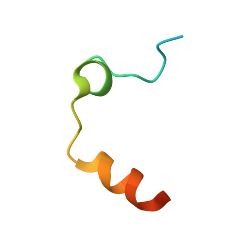
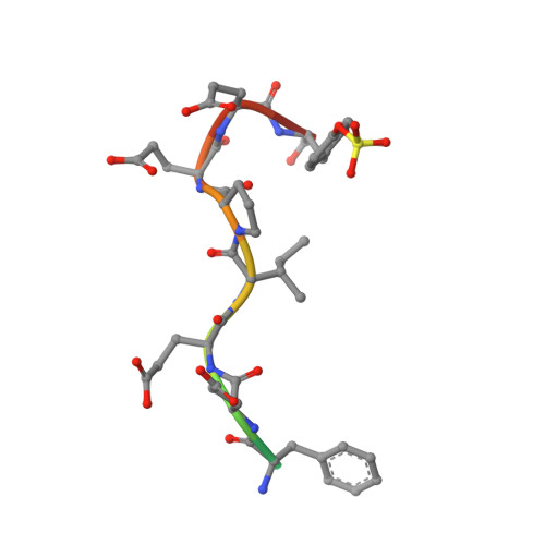
Hirudin variant-2C [auth I]12Hirudo medicinalisMutation(s): 0 
UniProt
Find proteins for P09945 (Hirudo medicinalis)
Explore P09945 
Go to UniProtKB:  P09945
Entity Groups  
Sequence Clusters30% Identity50% Identity70% Identity90% Identity95% Identity100% Identity
UniProt GroupP09945
Sequence Annotations
Expand
  • Reference Sequence
Small Molecules
Ligands 2 Unique
IDChains Name / Formula / InChI Key2D Diagram3D Interactions
2OJ (Subject of Investigation/LOI)
Query on 2OJ

Download Ideal Coordinates CCD File 
J [auth H]N-(2-{[5-(5-chlorothiophen-2-yl)-1,2-oxazol-3-yl]methoxy}-6-{3-[(2,3,4,6-tetra-O-acetyl-beta-D-glucopyranosyl)oxy]propoxy}phenyl)-1-(propan-2-yl)piperidine-4-carboxamide
C40 H50 Cl N3 O14 S
GNVQZTIXGHEXLF-NIURAOKHSA-N
DMS
Query on DMS

Download Ideal Coordinates CCD File 
D [auth H]
E [auth H]
F [auth H]
G [auth H]
H
DIMETHYL SULFOXIDE
C2 H6 O S
IAZDPXIOMUYVGZ-UHFFFAOYSA-N
Modified Residues  1 Unique
IDChains TypeFormula2D DiagramParent
TYS
Query on TYS
C [auth I]L-PEPTIDE LINKINGC9 H11 N O6 STYR
Binding Affinity Annotations 
IDSourceBinding Affinity
2OJ BindingDB:  6EO9 Ki: 160 (nM) from 1 assay(s)
Experimental Data & Validation

Experimental Data

  • Method: X-RAY DIFFRACTION
  • Resolution: 1.84 Å
  • R-Value Free:  0.248 (Depositor), 0.250 (DCC) 
  • R-Value Work:  0.198 (Depositor), 0.200 (DCC) 
  • R-Value Observed: 0.201 (Depositor) 
Space Group: C 1 2 1
Unit Cell:
Length ( Å )Angle ( ˚ )
a = 67.46α = 90
b = 71.64β = 100.21
c = 71.8γ = 90
Software Package:
Software NamePurpose
REFMACrefinement
MOSFLMdata reduction
SCALAdata scaling
Sir2014phasing

Structure Validation

View Full Validation Report


Ligand Structure Quality Assessment 

Created with Raphaël 2.3.0Worse 01 BetterLigand structure goodness of fit to experimental dataBest fitted 2OJClick on this verticalbar to view details

Entry History 

Deposition Data

Revision History  (Full details and data files)

  • Version 1.0: 2017-12-13
    Type: Initial release
  • Version 1.1: 2018-03-07
    Changes: Source and taxonomy
  • Version 1.2: 2019-10-16
    Changes: Data collection
  • Version 1.3: 2024-11-20
    Changes: Data collection, Database references, Structure summary