6ELU | pdb_00006elu

Structure of Serum Resistance Associated protein from T. b. rhodesiense


Experimental Data Snapshot

  • Method: X-RAY DIFFRACTION
  • Resolution: 2.30 Å
  • R-Value Free: 
    0.220 (Depositor), 0.222 (DCC) 
  • R-Value Work: 
    0.191 (Depositor), 0.192 (DCC) 
  • R-Value Observed: 
    0.192 (Depositor) 

wwPDB Validation 3D Report Full Report

Validation slider image for 6ELU

This is version 1.4 of the entry. See complete history

Literature

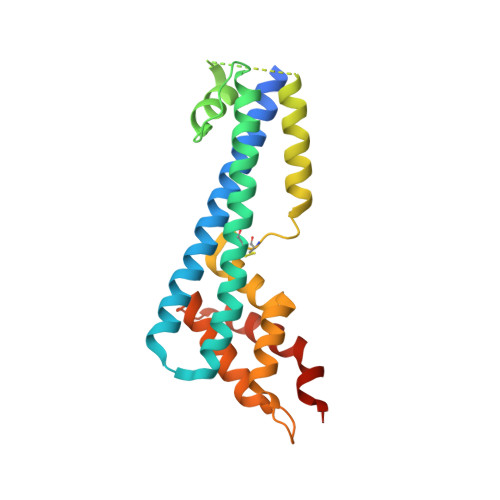
The structure of serum resistance-associated protein and its implications for human African trypanosomiasis.

Zoll, S.Lane-Serff, H.Mehmood, S.Schneider, J.Robinson, C.V.Carrington, M.Higgins, M.K.

(2018) Nat Microbiol 3: 295-301

  • DOI: https://doi.org/10.1038/s41564-017-0085-3
  • Primary Citation Related Structures: 
    6ELU

  • PubMed Abstract: 

    Only two trypanosome subspecies are able to cause human African trypanosomiasis. To establish an infection in human blood, they must overcome the innate immune system by resisting the toxic effects of trypanolytic factor 1 and trypanolytic factor 2 (refs.  1,2 ). These lipoprotein complexes contain an active, pore-forming component, apolipoprotein L1 (ApoL1), that causes trypanosome cell death 3 . One of the two human-infective subspecies, Trypanosoma brucei rhodesiense, differs from non-infective trypanosomes solely by the presence of the serum resistance-associated protein, which binds directly to ApoL1 and blocks its pore-forming capacity 3-5 . Since this interaction is the single critical event that renders T. b. rhodesiense human- infective, detailed structural information that allows identification of binding determinants is crucial to understand immune escape by the parasite. Here, we present the structure of serum resistance-associated protein and reveal the adaptations that occurred as it diverged from other trypanosome surface molecules to neutralize ApoL1. We also present our mapping of residues important for ApoL1 binding, giving molecular insight into this interaction at the heart of human sleeping sickness.


  • Organizational Affiliation
    • Department of Biochemistry, University of Oxford, Oxford, UK.

Macromolecule Content 

  • Total Structure Weight: 292.15 kDa 
  • Atom Count: 19,153 
  • Modeled Residue Count: 2,450 
  • Deposited Residue Count: 2,716 
  • Unique protein chains: 3

Macromolecules

Find similar proteins by:|  3D Structure
Entity ID: 1
MoleculeChains  Sequence LengthOrganismDetailsImage
Serum resistance associated; VSG protein
A, D, G, J
233Trypanosoma brucei rhodesienseMutation(s): 0 
Gene Names: SRA
UniProt
Find proteins for Q8T309 (Trypanosoma brucei rhodesiense)
Explore Q8T309 
Go to UniProtKB:  Q8T309
Entity Groups
Sequence Clusters30% Identity50% Identity70% Identity90% Identity95% Identity100% Identity
UniProt GroupQ8T309
Sequence Annotations
Expand
Reference Sequence
Find similar proteins by:|  3D Structure
Entity ID: 2
MoleculeChains  Sequence LengthOrganismDetailsImage
G10_3 heavy chain
B, E, H, K
229Mus musculusMutation(s): 0 
Find similar proteins by:|  3D Structure
Entity ID: 3
MoleculeChains  Sequence LengthOrganismDetailsImage
G10_3 Light chain
C, F, I, L
217Mus musculusMutation(s): 0 

Experimental Data & Validation

Experimental Data

  • Method: X-RAY DIFFRACTION
  • Resolution: 2.30 Å
  • R-Value Free:  0.220 (Depositor), 0.222 (DCC) 
  • R-Value Work:  0.191 (Depositor), 0.192 (DCC) 
  • R-Value Observed: 0.192 (Depositor) 
Space Group: P 1 21 1
Unit Cell:
Length ( Å )Angle ( ˚ )
a = 127.6α = 90
b = 103.55β = 114.58
c = 150.64γ = 90
Software Package:
Software NamePurpose
PHENIXrefinement
XDSdata reduction
XDSdata scaling
PHASERphasing

Structure Validation

View Full Validation Report


Entry History 

& Funding Information

Deposition Data


Funding OrganizationLocationGrant Number
Medical Research Council (United Kingdom)United KingdomMR/P001424/1

Revision History  (Full details and data files)

  • Version 1.0: 2018-01-24
    Type: Initial release
  • Version 1.1: 2018-01-31
    Changes: Database references
  • Version 1.2: 2018-02-28
    Changes: Database references
  • Version 1.3: 2019-10-16
    Changes: Data collection
  • Version 1.4: 2024-10-23
    Changes: Data collection, Database references, Structure summary