6EIH

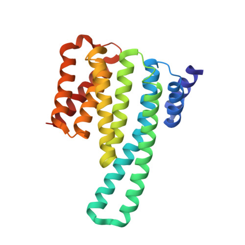
The crystal structure of 14-3-3 epsilon in complex with the phosphorylated NELFE peptide


Experimental Data Snapshot

  • Method: X-RAY DIFFRACTION
  • Resolution: 2.70 Å
  • R-Value Free: 0.241 
  • R-Value Work: 0.172 
  • R-Value Observed: 0.179 

wwPDB Validation   3D Report Full Report


This is version 1.5 of the entry. See complete history


Literature

p38-MK2 signaling axis regulates RNA metabolism after UV-light-induced DNA damage.

Borisova, M.E.Voigt, A.Tollenaere, M.A.X.Sahu, S.K.Juretschke, T.Kreim, N.Mailand, N.Choudhary, C.Bekker-Jensen, S.Akutsu, M.Wagner, S.A.Beli, P.

(2018) Nat Commun 9: 1017-1017

  • DOI: https://doi.org/10.1038/s41467-018-03417-3
  • Primary Citation of Related Structures:  
    6EIH

  • PubMed Abstract: 

    Ultraviolet (UV) light radiation induces the formation of bulky photoproducts in the DNA that globally affect transcription and splicing. However, the signaling pathways and mechanisms that link UV-light-induced DNA damage to changes in RNA metabolism remain poorly understood. Here we employ quantitative phosphoproteomics and protein kinase inhibition to provide a systems view on protein phosphorylation patterns induced by UV light and uncover the dependencies of phosphorylation events on the canonical DNA damage signaling by ATM/ATR and the p38 MAP kinase pathway. We identify RNA-binding proteins as primary substrates and 14-3-3 as direct readers of p38-MK2-dependent phosphorylation induced by UV light. Mechanistically, we show that MK2 phosphorylates the RNA-binding subunit of the NELF complex NELFE on Serine 115. NELFE phosphorylation promotes the recruitment of 14-3-3 and rapid dissociation of the NELF complex from chromatin, which is accompanied by RNA polymerase II elongation.


  • Organizational Affiliation

    Institute of Molecular Biology (IMB), Ackermannweg 4, 55128, Mainz, Germany.


Macromolecules
Find similar proteins by:  (by identity cutoff)  |  3D Structure
Entity ID: 1
MoleculeChains Sequence LengthOrganismDetailsImage
14-3-3 protein epsilon230Homo sapiensMutation(s): 0 
Gene Names: YWHAE
UniProt & NIH Common Fund Data Resources
Find proteins for P62258 (Homo sapiens)
Explore P62258 
Go to UniProtKB:  P62258
PHAROS:  P62258
GTEx:  ENSG00000108953 
Entity Groups  
Sequence Clusters30% Identity50% Identity70% Identity90% Identity95% Identity100% Identity
UniProt GroupP62258
Sequence Annotations
Expand
  • Reference Sequence

Find similar proteins by:  Sequence   |   3D Structure  

Entity ID: 2
MoleculeChains Sequence LengthOrganismDetailsImage
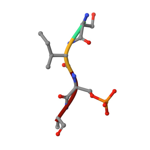
SER-ILE-SEP-ARGB [auth P]4Homo sapiensMutation(s): 0 
Sequence Annotations
Expand
  • Reference Sequence
Small Molecules
Modified Residues  1 Unique
IDChains TypeFormula2D DiagramParent
SEP
Query on SEP
B [auth P]L-PEPTIDE LINKINGC3 H8 N O6 PSER
Experimental Data & Validation

Experimental Data

  • Method: X-RAY DIFFRACTION
  • Resolution: 2.70 Å
  • R-Value Free: 0.241 
  • R-Value Work: 0.172 
  • R-Value Observed: 0.179 
  • Space Group: C 2 2 21
Unit Cell:
Length ( Å )Angle ( ˚ )
a = 79.29α = 90
b = 81.186β = 90
c = 81.05γ = 90
Software Package:
Software NamePurpose
SCALAdata scaling
MOLREPphasing
REFMACrefinement
PDB_EXTRACTdata extraction

Structure Validation

View Full Validation Report


Entry History & Funding Information

Deposition Data


Funding OrganizationLocationGrant Number
Emmy Noether ProgramGermany5342/1-1
SFBGermanySFB 1177
Marie Curie Career Integration Grant from the European Commission630763
LOEWE program Ubiquitin NetworksGermany--

Revision History  (Full details and data files)

  • Version 1.0: 2018-02-14
    Type: Initial release
  • Version 1.1: 2018-03-21
    Changes: Database references
  • Version 1.2: 2019-03-13
    Changes: Data collection, Derived calculations
  • Version 1.3: 2019-07-10
    Changes: Data collection
  • Version 1.4: 2019-10-16
    Changes: Data collection
  • Version 1.5: 2024-11-20
    Changes: Data collection, Database references, Structure summary